5 Operating procedures
32 GE Healthcare 86500-IMG rev 3
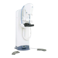
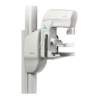
3 Slightly rotate the patient medially
and lean her towards the unit. Refer
to Fig. 5.10, Patient positioning at
cranio-caudal projection. Allow the
patient to grip the hand grips, and
lean towards the unit so that the
cassette holder under the breast
makes contact against the ribs.
4 Place the breast on the cassette
holder so that it is symmetrical and
totally covers at least one of the
automatic exposure control
detectors, as demonstrated by the
graphics printed on the clear acrylic
of the compression paddle.
Manually select the most appropriate detector or use the AutoPoint
automatic detector selection feature.
NOTE!
If the breast does not cover at least one detector or the breast has an
implant, a manually selected exposure technique must be used.
5 Apply the compression to the breast using the compression foot
pedal or the manual compression controls. Make sure that the nipple
is not inverted. Smooth out any skin wrinkles that may have been
caused during compression.
6 Position a film marker on the image receptor to the side of the lateral
aspect of the breast. Turn the patient’s head away from the X-ray
beam.
7 Take note of the compression thickness display to achieve the
correct compression for both breasts. When compressed correctly,
the breast should be firm when palpated. The skin may become
blushed, red or pink. Make sure that the ready light is on. Select or de-
select the Auto Rel (automatic compression release) function.
8 Make the exposure by pressing the exposure button, and keep it
pressed until the radiation indicator light goes out and the audible
exposure indicator stops. Release breast compression (if not
automatic).
9 Change the cassette and make an exposure of the other breast or
move on to another projection.
NOTE!
In manual usage, the selected kV should produce an exposure time from
0.5 to 1.2 seconds. If exposures are longer than 1.2 seconds, increase the
kV value. If they are shorter than 0.5sec., reduce the kV value.
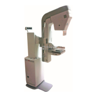
Fig 5.10. Patient positioning at
cranio-caudal projection
FOR TRAINING PURPOSES ONLY!
NOTE: Once downloaded, this document is UNCONTROLLED, and therefore may not be the latest revision. Always confirm revision status against a validated source (ie CDL).