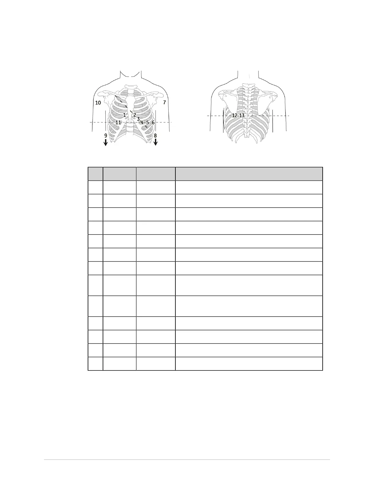
Patient Preparation
Table 47: Standard 15–Lead Electrode Placement
Item AHA Label IEC Label Description
1 V1 red C1 red Fourth intercostal space at the right sternal border.
2 V2 yellow. C2 yellow Fourth intercostal space at the left sternal border.
3 V3 green. C3 green Midway between location 2 and 4.
4 V4 blue C4 brown Mid-clavicular line in the fifth intercostal space.
5 V5 orange C5 black Anterior axillary line on the same horizontal level as 4.
6 V6 violet C6 violet Mid-axillary line on the same horizontal level as 4 and 5.
7 LA black L yellow Left deltoid.
8 LL red F green Above the left ankle (alternate placement— upper leg as close
to the torso as possible).
9 RL green N black Above the right ankle (alternate placement—upper leg as
close to the torso as possible).
10 RA white R red Right deltoid.
11 V4R gray C4R gray Right anterior chest opposite of 4.
12 V8 gray C8 gray Under left mid-scapular line.
13 V9 gray C9 gray Left paraspinal border.
Pediatric Electrode Placement
To acquire a pediatric ECG, use the electrode placement shown in the following
diagram.
146 MAC VU360
™
Resting ECG Analysis System 2088531-370-2