EasyManuals Logo

Mindray DC-80 User Manual

Mindray DC-80
383 pages
To Next Page IconTo Next Page
To Previous Page IconTo Previous Page
Page #184 background imageLoading...
Page #184 background image
5-108 Image Optimization
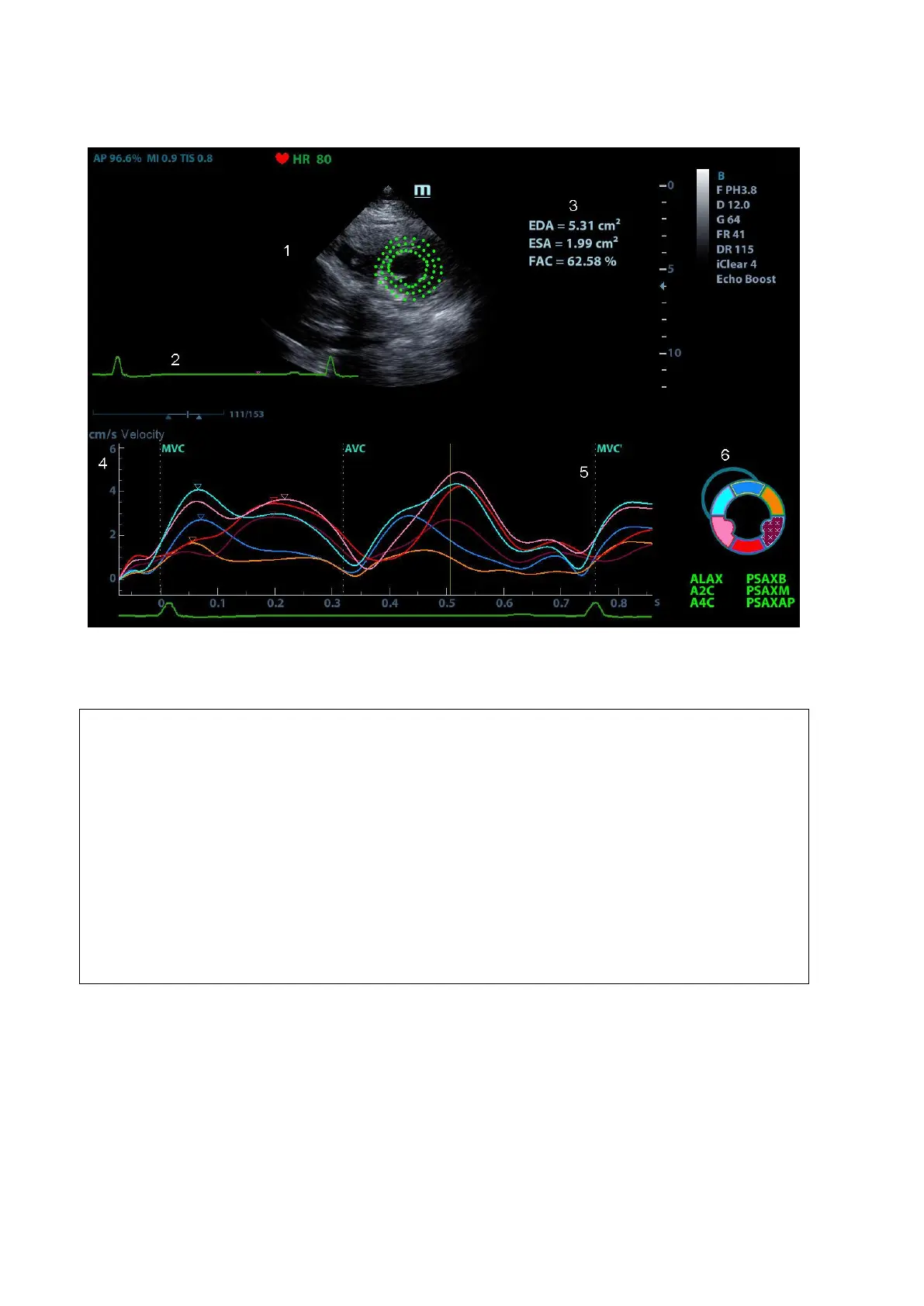
5.15.2 Tissue Tracking QA Screen Description
1Displays image used to generate trace curve
2Displays ECG trace
3Displays measurement and calculation results:
EDV:
maximum value of the end diastolic volume during the trace.
EDA:
maximum value of the end diastolic area (Left Ventricular) during the trace.
ESV:
maximum value of the end systolic volume (Left Ventricular) during the trace.
ESA:
maximum value of the end systolic area (Left Ventricular) during the trace.
FAC:
Fractional Area Change= (EDA ESA)/EDA.
This parameter is available under short axis section.
EF:
Ejection fraction.
This parameter is available under long axis section.
HR:
Heart rate.
GS:
Global Strain

Table of Contents

Questions and Answers:

Question and Answer IconNeed help?

Do you have a question about the Mindray DC-80 and is the answer not in the manual?

Mindray DC-80 Specifications

General IconGeneral
Imaging ModesB, M, Color Doppler, Power Doppler, Pulsed Wave Doppler, Continuous Wave Doppler, Tissue Doppler, 3D/4D Imaging
ProbesConvex, Linear, Phased Array, Volume
Advanced FeaturesAuto Optimization, Auto EF
ConnectivityDICOM 3.0, USB, HDMI, VGA, Ethernet
Transducer Ports4
ApplicationsAbdominal, Cardiac, Obstetrics/Gynecology, Vascular, Small Parts, Musculoskeletal, Urology, Pediatrics, Neonatal
Transducer TypesConvex, Linear, Phased Array, Volume
Power Supply100-240V AC, 50/60Hz
Operating Temperature10°C to 40°C
Storage Temperature-20°C to 60°C
Humidity30% to 80%

Related product manuals