EasyManuals Logo

Mindray DC-80 User Manual

Mindray DC-80
383 pages
To Next Page IconTo Next Page
To Previous Page IconTo Previous Page
Page #209 background imageLoading...
Page #209 background image
Image Optimization 5-133
3. Inject the contrast agent. Enable the timer, and save the dynamic image. See also Chapter
5.12 Contrast Imaging.
5.16.9 Parameter Setting
The parameters of the Fusion Imaging are described below:
Nav System
Operation
Tap [Tracking System] to enable or disable the function.
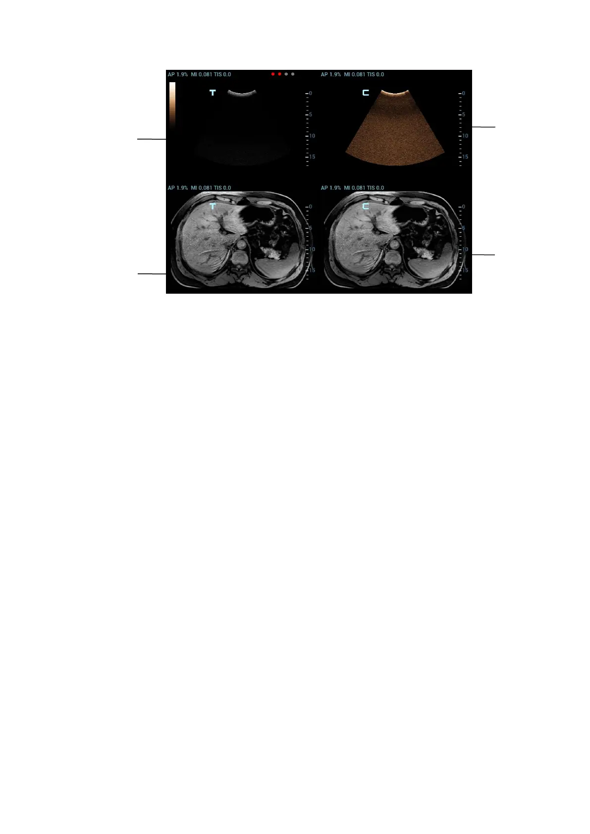
CT/MR image and the ultrasound image build up the coordinate relation. The
sensor indicator becomes green (navigation signal state indicator).
Fusion Ratio
Description
Adjust the display ratio that the ultrasound image registers with the CT/MR image.
Operation
Rotate the knob beneath [Fusion Ratio] to set the ratio.
The adjusting range is -1 ~ 1 in increments of 0.1.
0~1 represents that the ultrasound image registers with the CT/MR image; the
change of the fusion ratio appears on the right window side.
The larger the value is, the better the display effect becomes for the ultrasound
image registering with the CT/MR image; and vice versa. 0 only relates to
CT/MR image on the right window side and 1 only relates to ultrasound image
on the right window side.
-1~0 represents that the CT/MR image registers with the ultrasound image; the
change of the fusion ratio appears on the left window side.
The larger the value is, the poorer the display effect becomes for the CT/MR
image registering with the ultrasound image; vice versa. -1 only relates to
CT/MR image on the left window side and 0 only relates to ultrasound image on
the left window side.
1
2
3
4

Table of Contents

Questions and Answers:

Question and Answer IconNeed help?

Do you have a question about the Mindray DC-80 and is the answer not in the manual?

Mindray DC-80 Specifications

General IconGeneral
Imaging ModesB, M, Color Doppler, Power Doppler, Pulsed Wave Doppler, Continuous Wave Doppler, Tissue Doppler, 3D/4D Imaging
ProbesConvex, Linear, Phased Array, Volume
Advanced FeaturesAuto Optimization, Auto EF
ConnectivityDICOM 3.0, USB, HDMI, VGA, Ethernet
Transducer Ports4
ApplicationsAbdominal, Cardiac, Obstetrics/Gynecology, Vascular, Small Parts, Musculoskeletal, Urology, Pediatrics, Neonatal
Transducer TypesConvex, Linear, Phased Array, Volume
Power Supply100-240V AC, 50/60Hz
Operating Temperature10°C to 40°C
Storage Temperature-20°C to 60°C
Humidity30% to 80%

Related product manuals