3-18 System Preparation
Image area
The image area displays the ultrasound images, ECG waveforms, probe mark (or activating
window mark), time line (in M or PW mode), coordinate axis (including depth, time,
velocity/frequency), focal position (located at depth axis in the form of ), besides, the
annotation, body mark, measurement calipers, color bar/grayscale bar are also display here.
Parameter area
Displays the image parameters for the active window. If there are more than one imaging
modes, the parameters are displayed by each mode.
Cine Review area
Displays the cine review progress bar to indicate the cine replay progress.
ECG curve
ECG Display
Tip: amplitude and position of ECG waveforms can be changed.
Help information area
The help information area displays various help information or progress bar in the current
status.
Tips: in terms of help information, ―TB‖ refers to ―Trackball‖, ―Knob‖ refers to ―Multifunction
knob‖.
Press <Help> to open the Help Info manual.
Thumbnail
Displays the thumbnail images stored under the current patient.
Grayscale/ color bar
Displays the grayscale/ color bar corresponding to the current mode.
Soft Menu area
The monitor soft menu area displays the items that simultaneously appear at the bottom of the
touch screen.
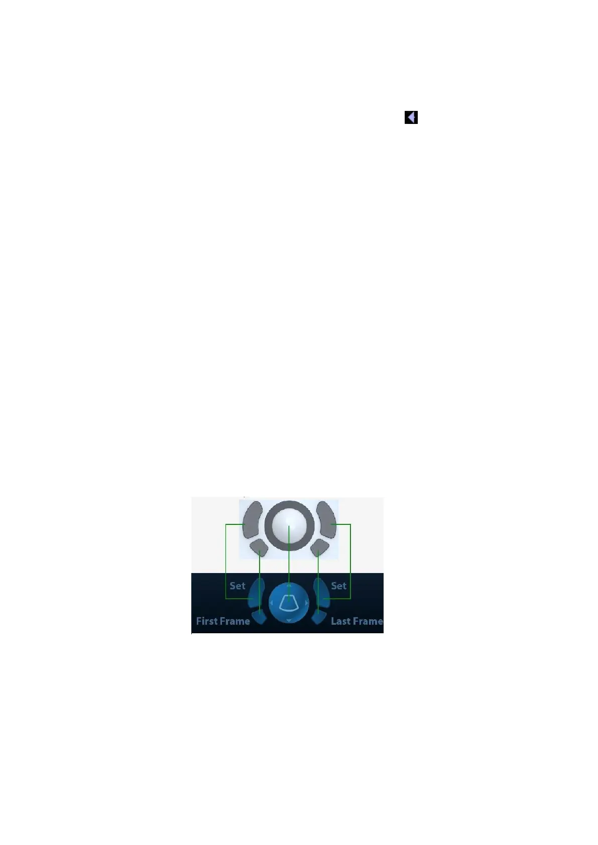
Trackball function hint area
Displays the current functions of trackball and keys, as shown in the following figure:
The left/right kidney shaped key is the <Set> key, to confirm the operation, similar to the left-
button of the mouse. Press to confirm an operation, same as the left-button of a mouse.
System status icon
This area displays the relevant system icons, such as USB memory device, printer, network,
Chinese / English entry, and current system time, etc.
Other Operations
Position of areas illustrated here are not fixed, you can move them by the trackball within a
certain area on the monitor.
Result window