Siemens AG SPB7-250.815.06.02.02 MAMMOMAT Novation DR
12.05 CS SD 24
Collimator Adjustment 51
Page 51 of 116
Medical Solutions
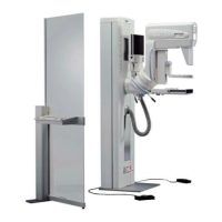
4. Select
Configuration -> Collimator -> Calibration of beam field
Fig. 33: Collimator beam calibration
5. Enter the numbers in the table below so that you always know what settings were used
in the beginning:
Tab. 10 Collimator beam calibration
6. Narrow the X-ray field so that the edges of the detector will not be exposed to X-ray and
white lines are visible in the image at the edges of the detector. To do so, decrease the
YC value and increase the YS, XR and XL values by 5 numbers.
7. Select a focus & anode combination.
8. Make sure a patient is registered and start an exposure.
9. Use a magnification of 4 in the application software and go to the edges of the detector.
White areas should be visible at all edges of the image. This shows you that the X-ray
does not cover the entire detector and shows where the beam really ends.
10. Measure the white line thickness at each image edge with the application GUI and ad-
just the corresponding XL, XR, YC and YS values with the service software.
X 11. Start an exposure.
12. Check if the while lines at the edges of the image are gone, using a magnification of 4 in
the application graphical user interface.
Focus and Anode XL XR YC YS
Small focus molly
Large focus molly
Small focus tungsten
Large focus tungsten
 Loading...