89
GENERAL INFORMATION ON USAGE AND MAINTENANCE
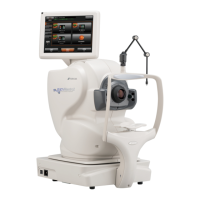
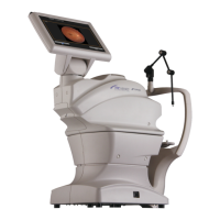
OPERATION PRINCIPLE
The patient's eye is illuminated by infrared light, which is emitted by the illumination optical system (IR
LED). The fundus observation/photography optical system forms an image on the fundus observation/
photography camera, and the image is observed on the control panel.
The auto alignment function works as follows: the anterior segment of the patient is illuminated by
infrared light emitted by the anterior segment observation LED. The auto alignment optical system
detects the pupil position with the anterior segment observation camera (master/slave). Then, the
instrument is moved to a position applicable to photography.
The auto focus function works as follows: the split lines (infrared light) emitted by the split LED are pro-
jected to fundus and the reflected image is detected by the fundus observation/photography camera.
Then, arithmetic processing is performed for the reflected image. The lens in the observation/photog-
raphy optical system is moved to a proper position by the auto focus mechanism (TRC focusing sensor
and TRC focusing motor) to adjust focus correctly.
The auto shoot function works as follows: when the capturing position and focus are applicable to pho-
tography by the auto alignment/focus mechanisms, the photography light source (xenon lamp) emits
light and the fundus observation/photography camera takes a picture.
The captured image can be recorded as electronic data by a personal computer or a commercial mem-
ory device connected to the external input/output terminal and can be printed by a printer.
It is possible to set the following functions for the image when the center (equivalent to picture angle
30°) of the captured image is extracted by software:
• Digital mask: The digital mask displays and saves the above-mentioned extracted image.
• Digital zoom: The above-mentioned extracted image is enlarged and the enlarged image is displayed
and saved by the digital zoom function.
In fundus stereoscopic photography, the instrument changes the captured positions (temporal side and
nasal side of pupil) and takes two pictures of one eye.
In fundus panorama photography, the instrument guides the patient's eye by the peripheral fixation tar-
get and takes a picture of the fundus periphery.
CHECKPOINTS FOR MAINTENANCE
1. Regularly maintain and check the instrument and its parts.
2. When using the instrument after a prolonged period of inactivity, confirm normal and safe operation
beforehand.
3. To take a good picture, be careful not to stain the objective lens with fingerprints or dust.
4. When this instrument is not in use, cap the objective lens and cover the instrument with the dust
cover.
5. When the objective lens is stained, clean it according to "Cleaning the objective lens" in this manual.