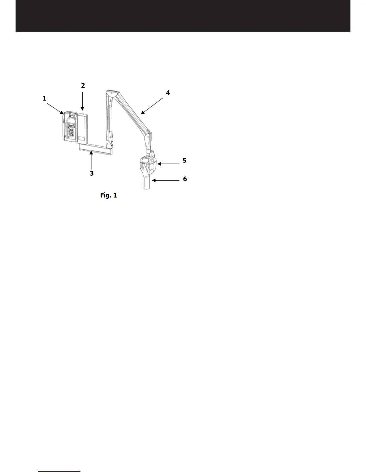
2.2. SYSTEM COMPONENTS
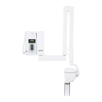
“Xmind®dc” radiographic system (Fig. 1) consists of:
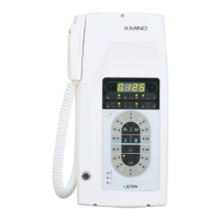
1. “Xmind®dc” TIMER
The timer is the control panel used to manage the exposure times and to safely use the tubehead.
To make the exposure, the control button with safety key is available.
The timer can be connected to n° 2 dc tubeheads.
2. WALL PLATE
3. BRACKET
The horizontal bracket is available in 3 different lengths (110 cm, 80 cm, 40 cm) and represents the support for
the pantograph arm. Its shaft is xed in a dedicated section of the timer (top or bottom) and allows for 180°
movement.
4. PANTOGRAPH ARM
Thanks to the new shape and new mechanisms of the positioning arm, it can be adjusted in height and depth
in order to precisely explore any spot in its reach.
It is made of light alloy with an ABS coating.
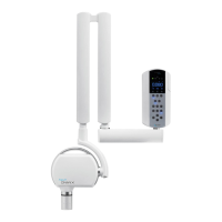
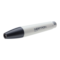
5. TUBEHEAD “Xmind®dc”
The intra-oral “Xmind®dc” is a tubehead type and its light alloy housing is divided into two compartments.
The high voltage transformer, the X-ray tube and the expansion chamber are submerged in highly dielectric
insulating oil inside a light alloy container.
The expansion chamber guarantees an adequate compensation to oil expansion for the entire temperature
range.
The X-ray tube is located in the back part of the container, allowing a source-skin distance 50% higher than
traditional structures.
In the second compartment the main electronic board and the control electronic board are placed.
6. CONE
The collimator cone or Beam Limiting Device represents the applied part of the device. Made of the transparent
polycarbonate, it allows:
- the correct distance between focal spot and skin
- dimension, direction and centering of X-ray beam
- the realization of different radiographic technique (bisecting and parallel technique).
During X-ray exposition, the collimator cone comes in contact with the skin of the patient. Before each exam, it is