Image Optimization 5-41
5.10.2 Note before Use
5.10.2.1 3D/4D Image Quality Conditions
In accordance with the ALARA (As Low As Reasonably Achievable) principle, please try
to shorten the sweeping time after a good 3D imaging is obtained.
The quality of images rendered in the 3D/4D mode is closely related to the fetal condition, angle of
a B tangent plane and scanning technique (only for Smart 3D). The following description uses the
fetal face imaging as an example, the other parts imaging are as the same.
Fetal Condition
(1) Gestational age
Fetuses of 24~30 weeks old are the most appropriate for 3D imaging.
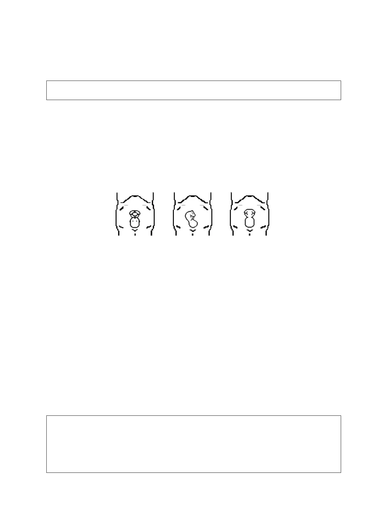
(2) Fetal body posture
Recommended: cephalic face up (Figure a) or face aside (Figure b);
NOT recommended: cephalic face down (Figure c).
(3) Amniotic fluid(AF) isolation
The region desired is isolated by amniotic fluid adequately.
The region to imaging is not covered by limbs or umbilical cord.
(4) The fetus keeps still. If there is a fetal movement, you need a rescanning.
Angle of a B tangent plane
The optimum tangent plane to the fetal face 3D/4D imaging is the sagittal section of the face.
To ensure high image quality, you’d better scan maximum face area and keep edge continuity.
Image quality in B mode (2D image quality)
Before entering 3D/4D capture, optimize the B mode image to assure:
High contrast between the desired region and the AF surrounded.
Clear boundary of the desired region.
Low noise of the AF area.
Scanning technique (only for Smart 3D)
Stability: body, arm and wrist must move smoothly, otherwise the restructured 3D image
distorts.
Slowness: move or rotate the probe slowly.
Evenness: move or rotate the probe at a steady speed or rate.
1. A region with qualified image in B mode may not be optimal for 3D/4D imaging. E.g.
adequate AF isolation for one MPR doesn’t mean the whole desired region is
isolated by AF.
2. More practices are needed for a high success rate of qualified 3D/4D imaging.
3. Even with good fetal condition, to acquire an approving 3D/4D image may need
more than one scanning.