5-44 Image Optimization
Function: set Min as 3D image rendering mode, displays the
minimum echo intensity in the observation direction.
This is helpful for viewing vessels and hollow structures.
Function: set X-ray as 3D image rendering mode. Displays the
average value of all gray values in the ROI.
X Ray: used for imaging tissues with different structure inside
or tissues with tumor.
Function: add the light rendering effect based on the general
rendering effect. It supports the global illumination and local
scattering.
5.10.3.3 Static 3D Image Viewing
Enter/Exit Image Viewing
To enter image viewing:
The system enters image viewing when image acquisition is finished.
Exit
To return to 3D/4D image acquisition preparation status, you can,
Press <Update> or <Freeze>.
Activate MPR
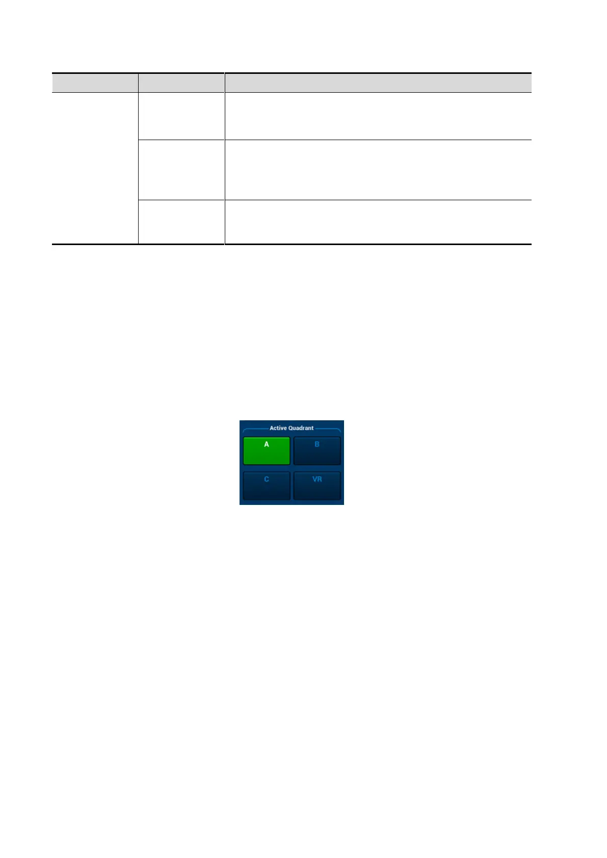
Touch [A], [B], [C] or [VR] to activate MPR or 3D image (VR).
MPR Viewing
In actual display, different colors of the window box and the section line are used to identify the
MPR A, B and C.
The color of window A is blue, and the color of the lines (representing MPR A) displayed in
the other two windows is blue as well.
The color of window B is yellow, and the color of the lines (representing MPR B) displayed
in the other two windows is yellow as well.
The color of window C is orange, and the color of the lines (representing MPR C)
displayed in the other two windows is orange as well.
Positions of the other two MPRs are indicated in the selected plane. You can roll the track ball to
change the position.