Image Optimization 5-75
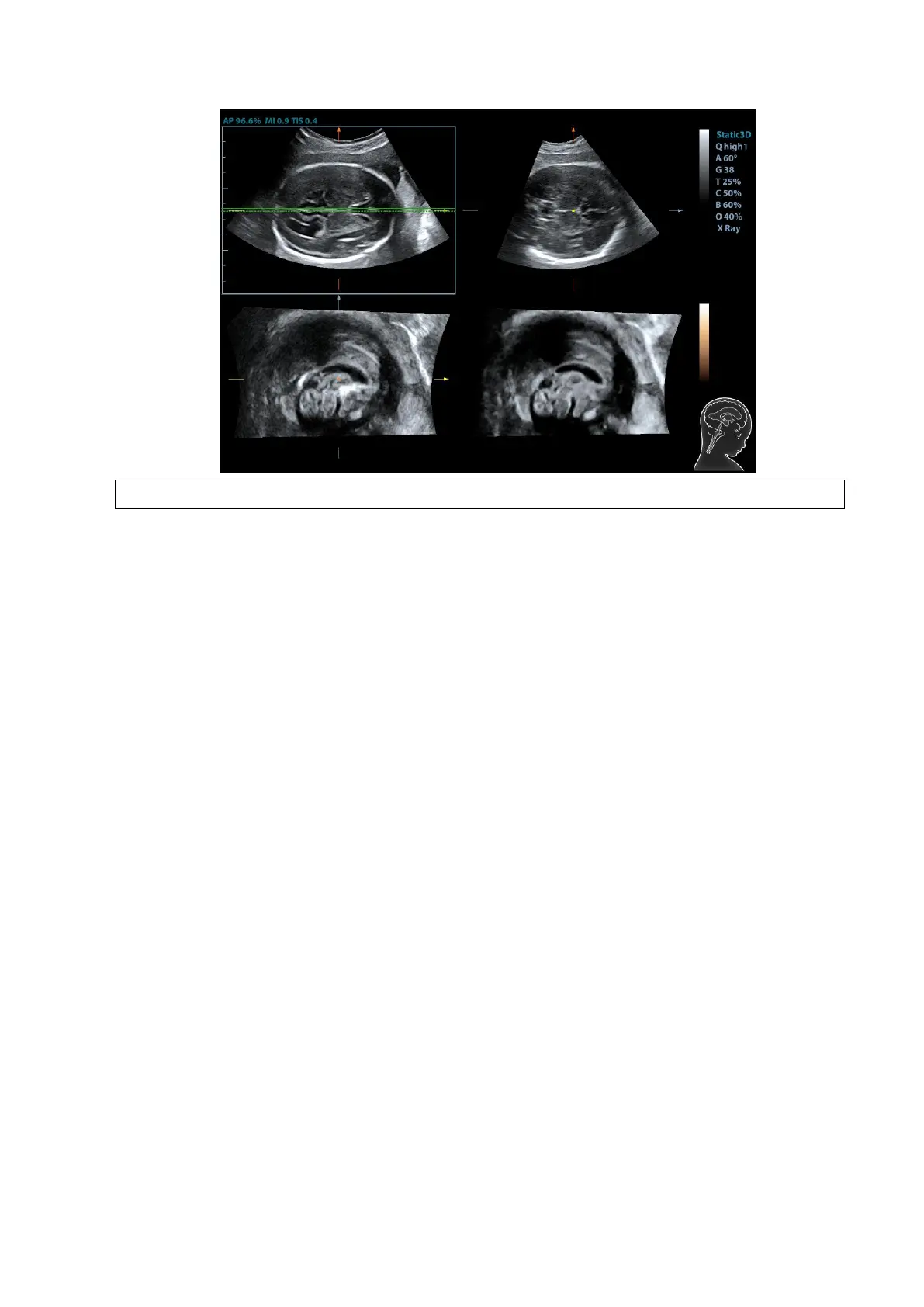
To ensure the correctness of the result, please select a clear sectional image.
5. Tap [OK] to accept the edit to the MSP. The system recalculates the TCP, TTP and TVP
according to MSP’s position. The position and the angle for TCP, TTP and TVP appears on
MSP plane.
6. Tap [TCP]/[TTP]/[TVP] to select the plane, and rotate <M>, <PW> or <C> knob to rotate the
image plane along with X/Y/Z axis. The angle value appears on the right of the image.
7. Rotate the reference line on the MSP plane. See Chapter 5.10.14.2 Other Operations for
details.
8. Tap [Auto Measure]. Tap [Edit] to edit the measurements. See Chapter 5.10.14.2 Other
Operations for details.
9. Click [Auto Comment], the system adds the orientation and the organ comments to the desired
area according to the active ultrasound image. See Chapter 5.10.14.2 Other Operations for
details.
10. Add the comment and body mark on the plane. Perform the measurement, and save the
single frame/multi-frame image.
5.10.14.2 Other Operations
Parameter adjusting
Brightness
Adjusting the brightness of the images.
Rotate [Brightness] to adjust the parameter.
The adjusting range is: 0-100% in increment of 2%.
Thickness
Rotate [Thickness] to adjust the parameter.
The adjusting range is: 0-30 in increment of 1.
The bigger the value is, the more the thickness information
appears.
3D iClear