EasyManuals Logo

Mindray DC-80 User Manual

Mindray DC-80
383 pages
To Next Page IconTo Next Page
To Previous Page IconTo Previous Page
Page #207 background imageLoading...
Page #207 background image
Image Optimization 5-131
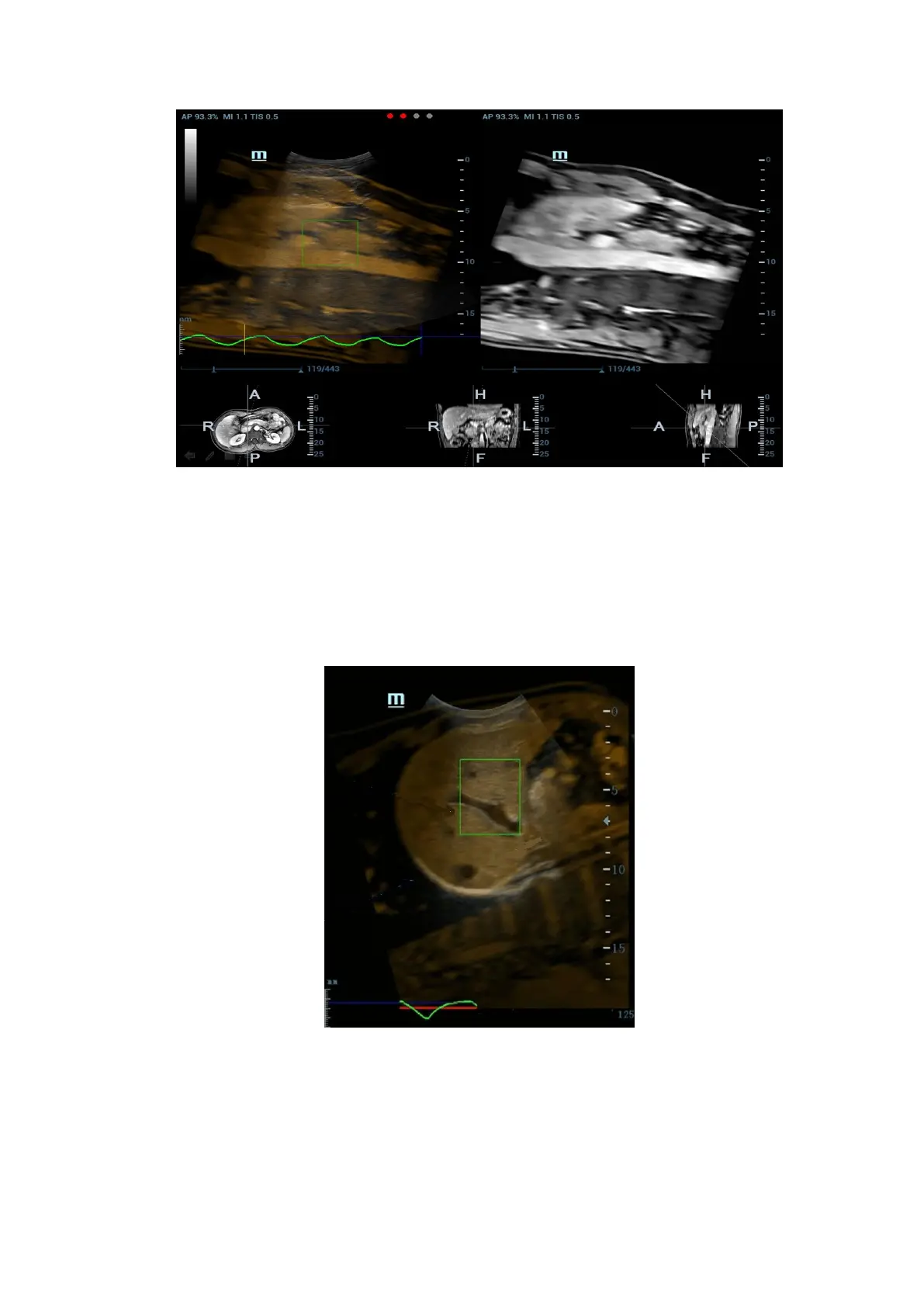
4. Tap [Motion Modeling]. If modelling succeeds, the system will play the cineloop automatically,
and ROI moves along with the motion of the respiration curve.
Note: RMQF scale is 0~1. 0 represents the poor motion modeling; 1 represents the premium
motion modeling.
Conduct step 4~5 repeatedly based on your demands. Set motion modeling repeatedly until a
premium one appears.
5. Tap [Motion Compen] to activate it. Move the probe. The Ultrasound System shows the CT
image which is processed by respiration compensation (Fusion Imaging with the respiration
compensation).
6. Save multi-frame cine.
Respiration Range
Description
The aspiration curve appears due to the active respiration depth. The respiration
curve beyond the scale becomes the straight line.

Table of Contents

Questions and Answers:

Question and Answer IconNeed help?

Do you have a question about the Mindray DC-80 and is the answer not in the manual?

Mindray DC-80 Specifications

General IconGeneral
Imaging ModesB, M, Color Doppler, Power Doppler, Pulsed Wave Doppler, Continuous Wave Doppler, Tissue Doppler, 3D/4D Imaging
ProbesConvex, Linear, Phased Array, Volume
Advanced FeaturesAuto Optimization, Auto EF
ConnectivityDICOM 3.0, USB, HDMI, VGA, Ethernet
Transducer Ports4
ApplicationsAbdominal, Cardiac, Obstetrics/Gynecology, Vascular, Small Parts, Musculoskeletal, Urology, Pediatrics, Neonatal
Transducer TypesConvex, Linear, Phased Array, Volume
Power Supply100-240V AC, 50/60Hz
Operating Temperature10°C to 40°C
Storage Temperature-20°C to 60°C
Humidity30% to 80%

Related product manuals