INTRODUCTION
Carl Zeiss Microscopy in transmitted-light brightfield in a few steps Axiovert 200
0-12 B 40-080 e 03/01
Microscopy in transmitted-light brightfield in a few steps
☞
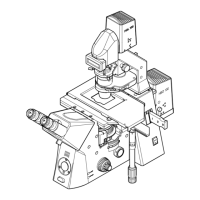
Before starting to use the Axiovert 200, make sure to read the notes on instrument safety and
the chapters entitled "Instrument Description" (Chapter 1) and "Start-up" (Chapter 2).
• Make the microscope ready for operation as described in chapter 2 and switch it on via the On/Off
switch (0-2/1).
• Select the objective with the lowest magnification (e.g. 10x) on the nosepiece (0-2/2). Set factor 1x on
the setting wheel (0-2/4) of the Optovar turret.
• Open the luminous-field diaphragm or the aperture diaphragm completely by pulling lever (0-2/16) to
the front until stop or by turning the setting wheel (0-2/20) to the front until stop.
• Turn the setting ring (0-2/19) to move the condenser turret in position H for brightfield (or DIC).
• Move reflector turret (0-2/5, if available) into the position without filter combination via the setting
ring.
• If required, remove analyzer slider (0-2/3) or switch to free light path.
• Turn setting wheel for Sideport right / left / vis (0-2/22) to position 100 % vis (visual
).
• Turn setting knob for Frontport / Baseport / vis (0-2/23) to position 100 % vis (
VIS
).
• Set beam splitting ratio to 100 % vis (0-2/10) on the tube. Switch off the Bertrand lens (if available).
Move combined rotary / slider knob (0-2/9) to position 100 % vis (
).
• Place a high-contrast specimen on the microscope stage (0-2/21). Adjust the binocular component.
• Use the coarse / fine focusing drive (0-2/6) to focus on the selected detail of the specimen. Should no
light be visible in the eyepieces, switch on the halogen illuminator via the HAL on / off switch (0-2/7).
• Use the toggle switch (0-2/8) to set the light
intensity to comfortable brightness.
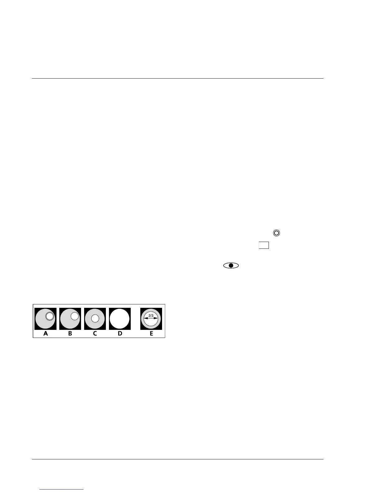
• Close luminous-field diaphragm (0-2/16) until it
is visible in the field of view, even if not in focus
(0-1/A).
• Focus on the edge of the luminous-field
diaphragm (0-1/B) by moving the condenser
(0-2/17) vertically.
• Center (0-1/C) luminous-field diaphragm via the centering screws (0-2/15 and 18) and open it until
the edge of the diaphragm just disappears from the field of view (0-1/D).
• Remove one eyepiece from the eyepiece tube (or swing in Bertrand lens) and set aperture diaphragm
(0-2/20) to approx. 2/3 of the diameter of the objective exit pupil (0-1/E). Optimum contrast setting is
dependent on the respective specimen.
• Insert the eyepiece again (or swing out Bertrand lens) and refocus, if required, via the fine drive.
• After the microscope has been set to transmitted-light brightfield in this way, changing to this special
contrasting technique is now possible (see chapter 3 of this manual).
Fig. 0-1 Diaphragm settings in transmitted-
light brightfield according to
KÖHLER