OPERATION
Axiovert 200 Contents Carl Zeiss
B 40-080 e 03/01 3-1
OPERATION
Contents
3 OPERATION...................................................................................................................3-3
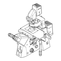
3.1 Axiovert 200 (manual) .....................................................................................................3-4
3.1.1 Operation and function controls on the Axiovert 200 (manual).........................................3-4
3.1.2 Switching on and basic settings on the Axiovert 200 (manual)........................................3-14
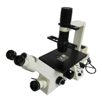
3.2 Axiovert 200 M (motorized)...........................................................................................3-16
3.2.1 Operation and function controls on the Axiovert 200 (motorized)...................................3-16
3.2.2 Switching on and basic settings on the Axiovert 200 M (motorized)................................3-23
3.3 Illumination and contrasting techniques .........................................................................3-28
3.3.1 Setting of transmitted-light brightfield for KÖHLER illumination......................................3-28
3.3.2 Setting of transmitted-light phase contrast.....................................................................3-33
3.3.3 Setting of differential interference contrast (DIC) in transmitted light..............................3-35
3.3.4 Setting of VAREL contrast in transmitted light ................................................................3-38
3.3.5 Setting of fluorescence contrast in reflected light ...........................................................3-40
3.4 Documentation..............................................................................................................3-43
3.4.1 Image orientation of camera ports.................................................................................3-43
3.4.2 Photomicrography with SLR camera...............................................................................3-45
3.4.3 Photomicrography using a digital camera and videomicroscopy ......................................3-46