䕔
1-8
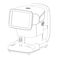
1.4 Overview
This instrument is designed to observe and analyze corneal endothelium by capturing an
image of corneal endothelium tissues without making contact, analyzing the captured
image, and calculating data such as cell density. In addition, this instrument also measures
the central corneal thickness while capturing an image of corneal endothelium tissues.
The patient places their chin on the chin rest and looks into the fixation light in the capture
window. The instrument automatically starts fine alignment when the physician roughly
aligns the corneal center and captures an image. An image can be captured manually
using the joystick if a problem with the eyes makes automatic operation impossible.
 Loading...