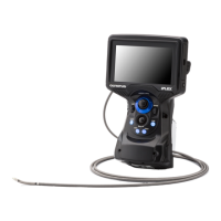
4-5 Observing the inspection object
1 Turn ON the illumination.
2 While looking at the display screen, insert the insertion tube into the inspection object.
Insert the insertion tube carefully while checking the insertion direction.
3 Use the [ANGLE/LOCK] joystick to perform angulation operations and observe the
applicable areas on the display screen.
Be careful not to apply the excessive pushing force, twisting or tension to the insertion tube.
4 Lock the angle and direction of the angulation section (angulation lock) and perform
observation.
When the angulation section is set to the desired angle and direction, press the [ANGLE/
LOCK] joystick.
The angulation angle can be adjusted finely even though the angulation is locked.
When the angulation is locked, the angle lock icon (
) appears in the upper right area of
the LCD monitor.
Adjust and record the image if necessary.
If the visibility of the inspection object deteriorates due to oil or other liquid stains, this may
be resolved by following the steps described in the notes below.
5 While looking at the display screen, pull out the insertion tube from the inspection
object slowly and carefully.
If the angulation is locked, press the [ANGLE/LOCK] joystick to unlock the angulation and
then, pull out the insertion tube.
NOTE
●
If the following message appears while inspecting, immediately stop observation,
carefully pull out the insertion tube, and perform the required action as instructed in
“Error messages” (page 97).
–
HIGH TEMPERATURE (DISTAL END). PLEASE IMMEDIATELY PULL OUT THE INSERTION
TUBE.
●
When operating the angulation section, do not bend the insertion tube to its minimum
bend radius or less (20 mm for 4 mm type, 30 mm for 6 mm type).
●
If the visibility of the inspection object deteriorates due to oil or other liquid stains when
using a forward-view optical adapter (excluding stereo optical adapters and the
AT100D/100S-IV76 optical adapter), this may be improved by operating the endoscope
such that the distal end comes into contact with the object to the left/right or in front of
the live image for 5 to 10 seconds. The degree of visibility may vary depending on the
type and amount of liquid stain.
41
 Loading...