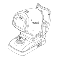
1-8
[Capture mode] : Ring topo mode and slit mode are available in this
instrument.
[RING TOPO mode] : Similar to conventional TMS, a mode to capture images of
the anterior shape of the cornea by projecting ring-shaped
light onto the cornea of the patient’s eye
[Slit mode] : Image of a corneal cross-section captured using the
Scheimpug principle in slit mode
[Mires ring. image] : Corneal image projected by multiple concentric rings of light
in ring topo mode
[Slit image] : Image of a corneal cross-section captured using the
Scheimpug principle in slit mode.
[Analysis screen] : There are two viewers (MERGED TOPO viewer and RING
TOPO viewer) with multiple analysis screens in each viewer.
[MERGED TOPO Viewer] : Displays the analysis screen that uses MERGED TOPO data
such as the MERGED TOPO map.
[RING TOPO Viewer] : Displays the analysis screen that uses RING TOPO data
such as the RING TOPO map.
[MERGED TOPO Map] : The cornea anterior shape map with analysis capability
enhanced by adding information obtained from images
captured in slit mode to the cornea anterior shape map
created from Mire ring images
[RING TOPO Map] : Cornea anterior shape map created from only Mire ring
images for the conventional TMS series, to be distinguished
from [MERGED TOPO Map]
[MERGED TOPO data] : Result of analysis conducted using both Mire ring images
and slit images.
[RING TOPO data] : Result of analysis conducted using only Mire ring images.
[Color map] : Curvature distribution map that shows the corneal shape
with contour lines.
 Loading...