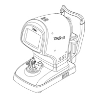
3-35
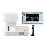
g) Slit Calculation
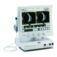
Measured slit images are listed. In addition, the analyzed
map and CCD image are also displayed as thumbnails.
Values vary depending on the specied measurement
point. Carefully examine the displayed value by
performing other examinations before use.
● Enlarging/reducing slit image
Click the “zoom” button (1) to increase or reduce the
image size. This function can also be selected from the
pull-down menu.
● Changing slit image
Click the “change slit image” button (2) to select the
slit image to be displayed. The slit image can also be
changed by dragging line (3) on the CCD image. Click
the “automatically change slit image” button (4) to
automatically play a slide show to the last slit image.
Click the “automatically change slit image” button (4)
again while playing a slide show to stop the automatic
play immediately.
● Analyzing slit image
If the anterior and posterior data of the cornea can be
analyzed on a slit image, click a measurement point on
(Fig. 1)
(19)
(7)
(10)
(6)
(16)
(18)
(12)
(11)
(3)
(8)
(1)
(2) (4)
(14)
(17)
(13)
(15)
(5)
(20)
(9)
(21)
(22)
 Loading...