䕔
3-18
- The image to be displayed immediately after automatic analysis can be
selected on the System Setup screen. Refer to “3.7.3 Application" for
setting method. The initial setting is “Area.”
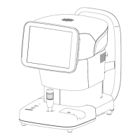
Ɣ Checking fixation light position
The position of the fixation light when capturing an image is shown on the R/L
eye analysis screen and single eye analysis screen with icons shown below.
- It can be set on the System Setup screen whether to indicate the position
for capturing an image with a black square on the icon. Refer to “3.7.3
Application" for setting method. The initial setting is OFF.
(1) Indication of fixation light position: OFF
Center
Parafo
va
30㼻
90㼻 150㼻 210㼻 270㼻
330㼻
Periph
eral
U
L.U L.L L R.L
R.U
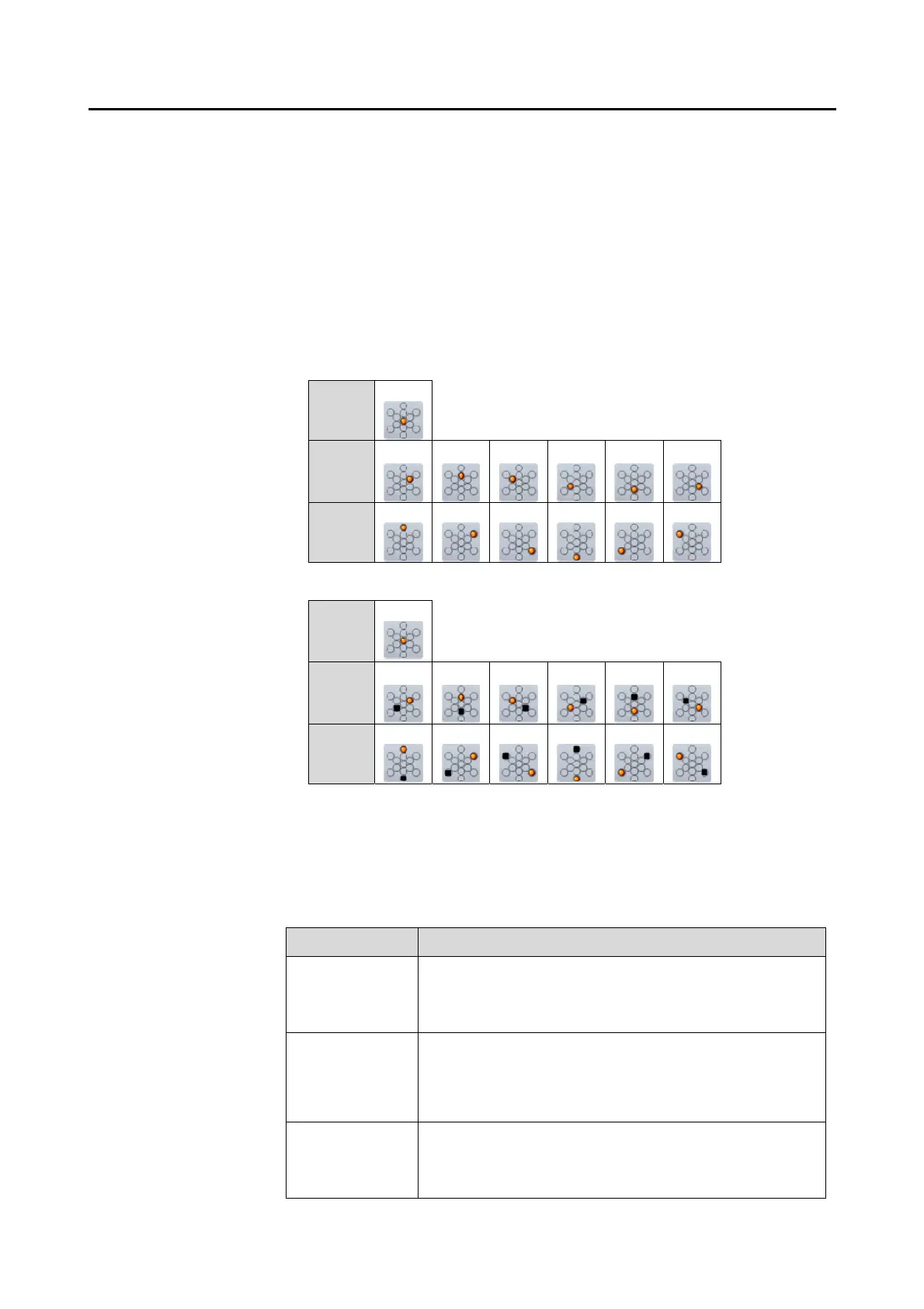
(2) Indication of fixation light position: ON
Center
Parafo
va
30㼻
90㼻 150㼻 210㼻 270㼻
330㼻
Periph
eral
U
L.U L.L L R.L
R.U
Ɣ Checking reliability
The displayed endothelium image is automatically analyzed and resultant
reliability is displayed on the R/L eye analysis screen and single eye analysis
screen. The reliability is categorized into 3 levels and indicated by the mark
corresponding to each level as listed below.
Reliability mark Description
No mark The reliability for automatic analysis is high because
cells are clearly captured over a wide range and cell
trace lines are extracted precisely by automatic
analysis.
e Because the range of captured cells is slightly less or
visibility of captured images is slightly poor, some cell
trace lines are not extracted correctly by automatic
analysis and the resultant reliability is slightly lower.
E Because the range of captured cells is small and
visibility of captured images is poor, cell trace lines are
not extracted correctly by automatic analysis and the
resultant reliability is low.
 Loading...