䕔
3-20
The following analysis results obtained by automatically extracting dark
areas from the displayed image of endothelium are displayed on the dark
area analysis screen (Fig. 4).
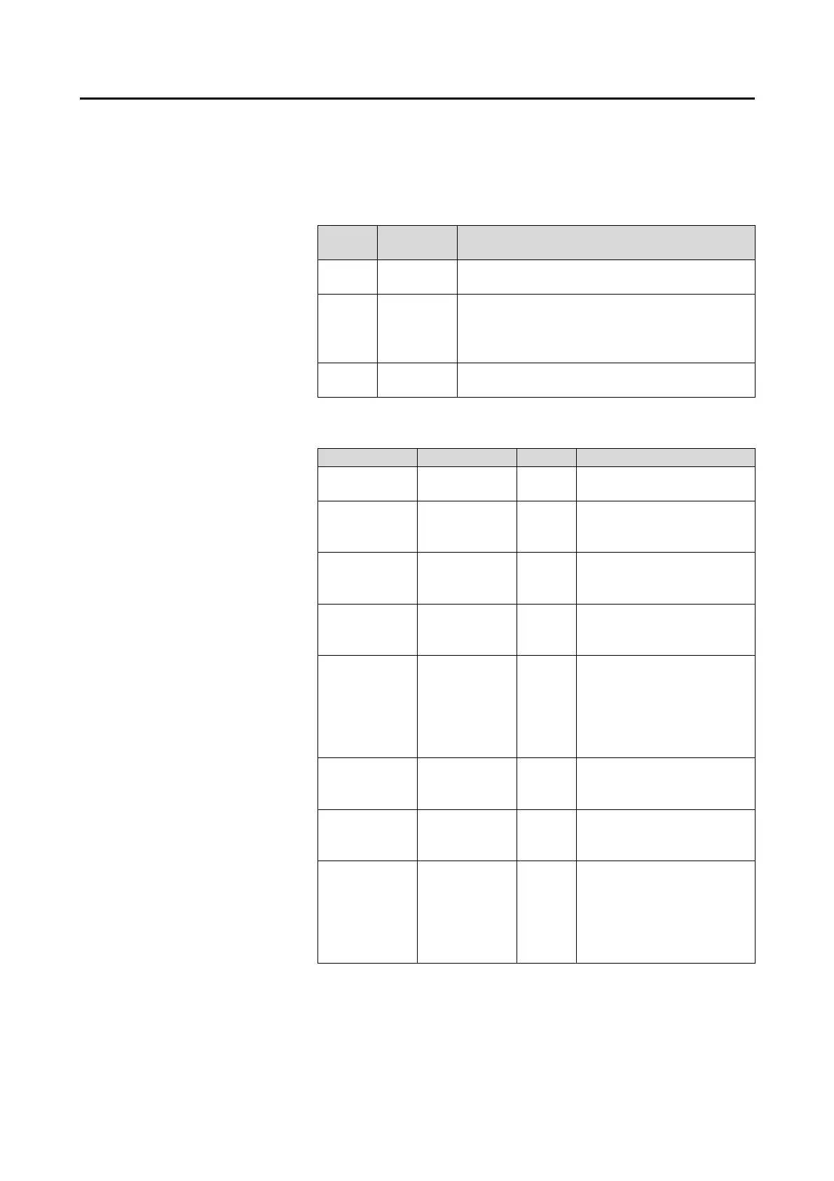
- Dark area analysis image
Use the following buttons to change the image.
Abbre
viation
Item Details
Photo Captured
image
Displays the captured image.
Trace Cell trace
line
Displays trace lines of extracted dark
areas..
Displays trace lines of endothelium tissue
in blue.
Area Area-spe
cific
Displays the extracted dark area
color-coded according to area sizes.
- Dark area analysis value
The analysis results of the displayed dark area in endothelium.
Abbreviation Item Unit Details
D.A.NUM Number of
dark areas
Pieces The number of analyzed
dark areas
D.A.Density Dark area
density
/mm
2
The number of cells in 1
mm
2
of the analyzed
dark area
D.A.AVG Average
area of dark
area
ȝm
2
Average area of
analyzed dark areas
D.A.SD Standard
deviation of
dark area
ȝm
2
Standard deviation of the
analyzed dark area
D.A.CV Fluctuation
coefficient to
dark area
% Fluctuation coefficient of
the analyzed dark area.
Value calculated by
dividing the standard
deviation with the
average area.
D.A.MAX Maximum
size of dark
area
ȝm
2
The area of the largest
cell in the analyzed dark
areas
D.A.MIN Minimum
size of dark
area
ȝm
2
The area of the smallest
cell in the analyzed dark
areas
D.A.Ratio Dark area
ratio
% Ratio of the dark area size
relative to the total area.
The total area is the sum of
analyzed dark area and the
area of analyzed
endothelium tissue.
- Distribution according to sizes of dark area
Distribution according to sizes of dark areas automatically extracted
is shown in a histogram.
 Loading...