4.3. POSITION OF THE X-RAY PLATE OR SENSOR
The parallel technique, where applicable, provides more accurate images in terms of size compared to the bisecting
technique. A rectangular collimator, with 30 cm (12"), focus-skin distance, is always preferable to obtain better quality
pictures. To avoid exposing the image receiver only partly (whether it is a sensor or photostimulable phosphorus
plate system) an alignment device that gives rectangular collimators guidelines should be used. These lines are
usually given on the alignment ring.
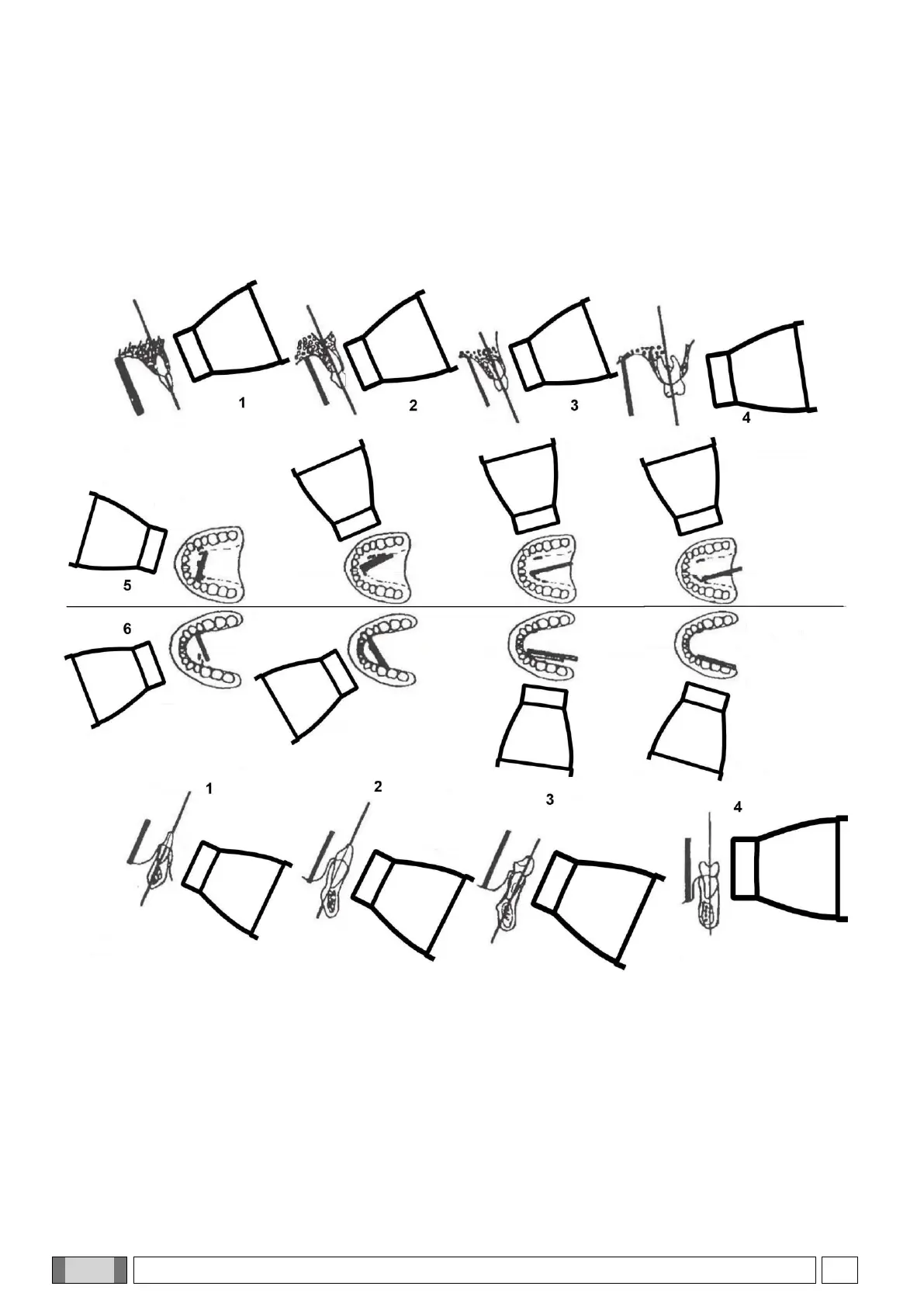
- Parallel technique.
- The x ray emission axis is perpendicular to the image receiver (for example a sensor or photostimulated
phosphor plate) which in turn is parallel with the tooth’s long axis.
- As a result, the picture of the tooth will only be deformed by the divergence of the x rays in relation to the focus
spot.
- Radiographic enlargement may reach up to 15%.
- For some “special” pictures, for example occluded ones, it may be necessary to remove the rectangular
collimator and use the round one if a positioner is not present.