Chapter 3 Preparation and Inspection
47
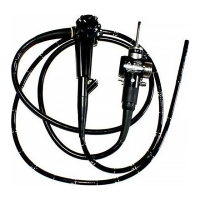
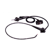
ULTRASOUND GASTROVIDEOSCOPE GF-UCT180
Figure 3.20
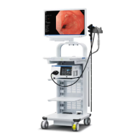
3.6 Inspection of the endoscopic system
Inspection of the endoscopic image
Do not stare directly into the distal end of the endoscope
while the examination light is ON. Otherwise, eye injury may
result.
1. Turn the video system center, light source, and monitor ON and inspect the
WLI and NBI endoscopic image as described in their respective instruction
manuals.
2. Confirm that light is output from the endoscope’s distal end.
3. While observing the palm of your hand, confirm that the WLI and NBI
endoscopic image is free from noise, blur, fog, or other irregularities.
4. Angulate the endoscope and confirm that the WLI and NBI endoscopic
image does not momentarily disappear or display any other irregularities.
If the object cannot be seen clearly, wipe the objective lens
using a clean, lint-free cloth moistened with 70% ethyl or
isopropyl alcohol.
Mark A (red)
Mark B (red)
Mark (red)
Mark A (red)
Mark B (red)
Mark (red)
Endoscope-side connector of the ultrasonic cable

 Loading...