Do you have a question about the Bruker ICON and is the answer not in the manual?
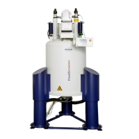
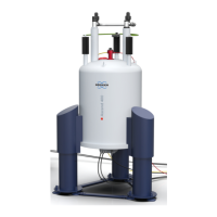
Details the ICON MRI system's shielded permanent 1 Tesla magnet and its features.
Explains the built-in gradient and shim coils, their cooling, and temperature supervision.
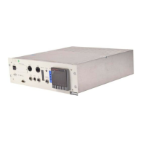
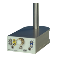
Describes the electronic cabinets housing spectrometer components for signal generation and detection.
Details the left-most cabinet prepared for Isofluorane evaporator and scavenging system.
Describes the table mounted on the anesthesia cabinet for animal preparation.
Overview of the modular animal support system including holder and exchangeable front pieces.
Details the animal holder with installed anesthesia, care, and supervision lines.
Describes various dedicated animal beds for mouse and rat examinations, including warming features.
Information on RF Coils pre-matched for in-vivo applications and their placement.
Describes tunable 1H single resonate transmit/receive RF Coils for animal beds.
Details RF Coils that can be mounted from the rear side of the instrument.
Information on the host computer acting as the operating console for the MR system.
Details the standard keyboard used with the MR system for operation.
Describes the two-button mouse with a wheeler used for system operation.
Overview of safety issues for system operation and references to other manuals.
Explains safety alert symbols, keywords, and notices used in the manual for hazard communication.
Provides instructions on shutting down the MR system using the ON/OFF switch.
Covers electrical safety, low voltage directive, and general hazard precautions.
Addresses hazards related to mechanical injury, particularly tripping over cables.
Discusses hazards from static, gradient, and radio frequency electromagnetic fields.
Details risks associated with the static magnetic field, including pacemaker interference and material attraction.
Explains risks from gradient fields, including peripheral nerve stimulation and burn injuries.
Covers hazards related to radio frequency fields, such as tissue heating and burn injuries.
Safety aspects concerning the use of RF Coils and accessories, including damage and leaks.
Safety precautions when using narcotic gases, including regulations and scavenging systems.
Guidelines for handling, spills, storage, and first aid for phantom fluids.
Safe handling practices for measurement phantoms to prevent personal injury.
Procedures for cleaning up phantom fluid spills and personal protection measures.
Recommendations for storing measurement phantoms to prevent fire and damage.
First aid measures for skin, eye, ingestion, and inhalation contact with phantom fluids.
Safety considerations for animals during MRI measurements, focusing on overheating and nerve stimulation.
Describes the procedures for powering the MRI system on, off, and the different operational states.
Lists and explains the four distinct operation states of the MRI scanner.
Step-by-step guide on how to switch the MRI system ON.
Step-by-step guide on how to switch the MRI system OFF.
Overview of the process for preparing and running MRI scans on the ICON system.
Introduction to the animal support system and the preparation steps for scans.
Outlines the general workflow for preparing and running scans using the ParaVision software.
Instructions on how to mount animal beds to the basic holder for scanning.
Provides a schematic layout and connection details for the mouse body animal bed.
Details the connections and features of the mouse body RF Coil.
Explains how to slide and fix the animal bed onto the basic holder.
Instructions on connecting RF Coils to the MRI system.
Details the connection of animal bed mounted RF Coil cables to the magnet.
Explains how to connect magnet mounted RF Coils using BNC cables.
Guidance on positioning the animal and setup within the magnet bore.
Step-by-step instructions for placing the animal correctly on the animal bed.
Instructions for placing the RF Coil on the animal bed for body imaging.
Procedure for smoothly inserting and fixing the animal setup into the magnet.
Explains the importance and process of tuning and matching RF Coils for optimal signal.
Workflow for tuning and matching the RF Coil inside or outside the magnet bore.
Details the general workflow for routine system operation, including selection and acquisition.
Provides a detailed workflow diagram for routine MRI scanning procedures.
Guidance on adapting scan protocols for mice and rats for routine work.
Recommended maximum scan times for different imaging methods.
Information on reachable spatial resolutions for anatomical imaging with the ICON system.
Details the programmable input and output triggers for scanner synchronization.
Explains the use of input trigger signals for synchronizing the scanner with external events.
Describes the use of output trigger signals to control or synchronize external devices.
Locates and labels the trigger connectors within the anesthesia cabinet.
Information on data storage and retrieval using the ParaVision Data Manager software.
Details the appropriate load phantoms for mouse and rat RF Coils and their usage.
Instructions on how to place phantoms on animal beds for reproducible results.
Overview of typical RF Coils and animal beds available for the MRI system.
Description of the optional MR compatible small animal life monitoring and gating system.
Information on the optional animal body warming unit connectable to ICON animal beds.
Guidance on performing regular checks to ensure system integrity and safety.
Lists essential checks on cooling fans, anesthesia system, and RF coils before each measurement.
Key daily checks, including the capacity of the scavenging filter.
Table outlining cleaning procedures for various system components like monitor and RF coils.
Provides a list of all tables included in the manual.
Provides a list of all figures included in the manual.
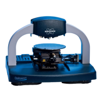
| Type | Atomic Force Microscope (AFM) |
|---|---|
| Manufacturer | Bruker |
| Sample Size | Up to 200 mm diameter |
| Scanning Range | 90 μm x 90 μm x 10 μm |
| Imaging Modes | Contact Mode, Tapping Mode, PeakForce Tapping |
| Operating Environment | Ambient |