Measurement and Analysis
Vivid S5/Vivid S6 User Manual 291
R2424458-100 Rev. 2
Peak detection
The peak systolic strain detection for each segment can be
verified and eventually manually changed.
To adjust the peak detection:
1. Press BE+Traces.
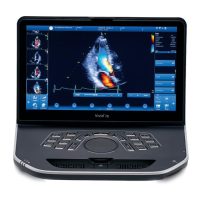
The Bulls-Eye and Traces screen is displayed
(Figure 7-21) showing:
• Trace plots for all three loops
• Bulls-Eye with Peak systolic strain values
2. To change the peak marker position on a curve:
• Press Set on the peak marker (square point) on one of
the curves, move the peak marker to a new position and
press the Set key again to fix the point.
- OR -
• Place the cursor on a segment in the Bulls-Eye. The
corresponding curve is highlighted.
3. Click on the segment to select the corresponding peak
marker and move it to a new position.
The position of the AVC marker can also be checked in the
Bulls-Eye and Traces screen. If needed, the APLAX view
should be reprocessed to change the AVC time.
About the Results
Be aware of the following:
• Clinical assessments should be made based on both color
and segmental Peak systolic strain values.
• The Save As function is intended for research purposes
and should not be used to archive diagnostic data.
• To populate the worksheet the report and the review page
the Single Bulls-Eye screen must be saved.
• All results shown (curves and colors) are based on drift
compensated values. Any strain drifting is linearly
compensated throughout the cycle. If the drift
compensation in a given segment is too high, the tracking
quality is automatically set to Not acceptable ( ).
• If the tracking quality was scored as Not acceptable
( ) in more than one segment, the Global peak strain
value is not calculated.
 Loading...