15-2
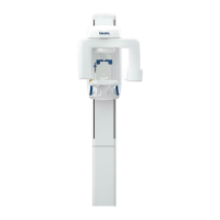
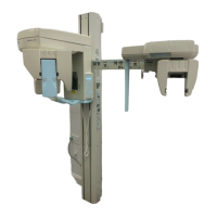
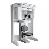
Gendex GXCB-500
TM
Service Manual
G990710 30 June 2008
Conditions of Operation – 8.9 and 23.0 Second Scans
The measurements were conducted by a certified health physicist from RayScan,
Inc, on the factory floor on 04.01.08. He was assisted by a physicist, employed by
the manufacturer.
The X-ray scatter was measured from a GXCB-500 Scanner in several modes,
using a Radcal Model 9010 Radiation monitor, with a 10X5-180 chamber.
A head phantom from Phantom Labs, Inc, was used as the scatter center and
placed in a representative location as measured by an image scan. Model SK150
has real bone as the structural component and proprietary Urethane filler that
simulates the response of tissue.
The radiation monitor was held at the same height as the phantom head’s nose and
the distances were measured from the center of rotation of the CT scan.
Measurements were taken every 45°, with 0° measured directly in front of the
scanner, and at distances of 3ft, 6ft, and 9ft. In addition the scatter was measured
at 3ft directly above the system and at a position 3ft above and 3ft directly in front
of the device.
Scatter was measured for the Standard (full-beam), Extended Diameter Scan
(EDS) (half-beam), and Panoramic mode. Both scan modes were taken with 0.4
mm Voxels for a scan time of 8.9s. The X-ray beam details were 120 kVp, 5 mA,
and 28.52 mAs. The Panoramic scan settings were measured with the Large
exposure setting with the following settings: 89kVp, 5 mA, and 71.43 mAs, 20s
scan time.
 Loading...