EasyManuals Logo

Mindray M7 User Manual

Mindray M7
361 pages
To Next Page IconTo Next Page
To Previous Page IconTo Previous Page
Page #136 background imageLoading...
Page #136 background image
5-56 Image Optimization
Procedures
(1) In image viewing mode, click [Accept VOI] to be “Off”.
(2) Select a desired sectional plane by clicking [Current Window X]. Then VOI on
section A, B and C are adjustable.
(3) Roll the trackball to change the ROI size and position as well as VOI curve, press
the <Set> key to toggle among setting the ROI size, position and VOI curve, or,
rotate the multifunction knob to change the position of VOI.
Click [Accept VOI] to be “On” to exit VOI adjusting. In Accept VOI, a green dotted-line
is displayed presenting the position of the section plane, using the trackball to move
the reference point on the MPR to slicing, and the point is the rotation center. Roll the
trackball to view a section image; you can see the corresponding MPR lines are
moving.
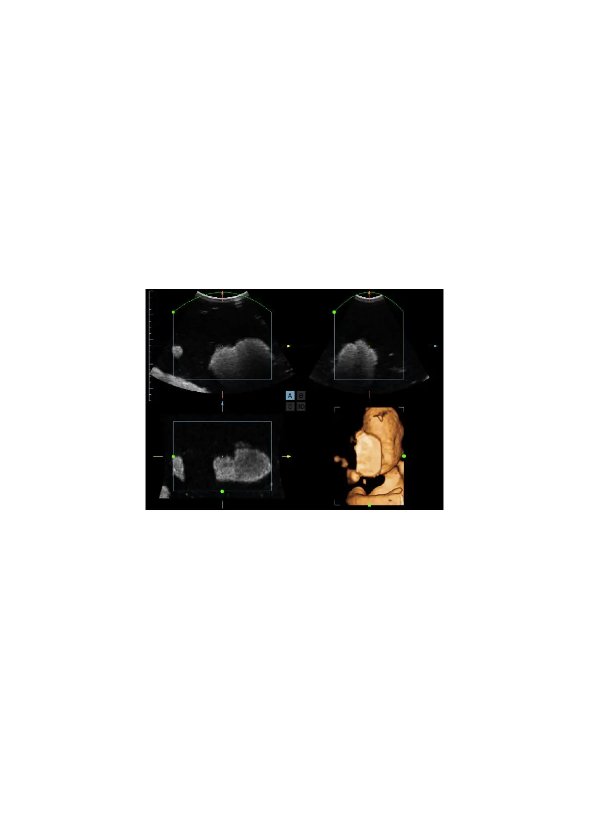
Figure a and Figure b below show the 3D image before and after VOI adjusting. VOI
adjusting helps to re-render a better 3D image.
a

Table of Contents

Other manuals for Mindray M7

Questions and Answers:

Question and Answer IconNeed help?

Do you have a question about the Mindray M7 and is the answer not in the manual?

Mindray M7 Specifications

General IconGeneral
TypePortable Ultrasound System
Display15-inch LCD
Imaging Modes2D, M-mode, Color Doppler, Power Doppler, Pulsed Wave Doppler, Continuous Wave Doppler
ProbesConvex, Linear, Phased Array
ApplicationsAbdominal, Cardiac, Vascular, OB/GYN, Small Parts, Musculoskeletal, Urology, Pediatric
BatteryRechargeable Lithium-ion
ConnectivityUSB, Ethernet, DICOM
Dimensions35 cm x 36 cm x 9 cm
Transducer Ports2 active transducer ports

Related product manuals