■
3-30
The following is not a requirement for capturing the measurement data, but a method
to confirm that the ultrasound wave correctly captures the geometrical axis and ideal
waveforms are taken.
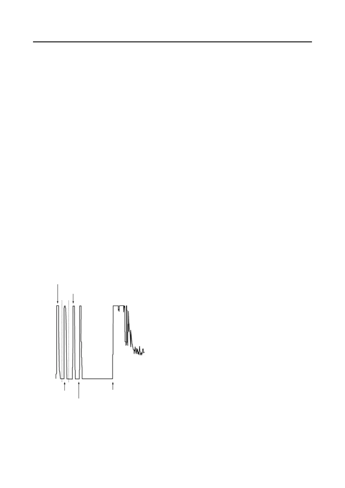
i. When the retina waveform spikes, the ultrasound wave reaches the retina
perpendicularly.
ii. When the waveforms on the front/back of the crystal lens spike, the ultrasound
wave correctly captures the geometrical axis.
iii. When the retina waveform can be distinguished from the sclera waveform, the
ultrasound wave reaches the retina perpendicularly. Because the depression
between two waveforms (choroid) cannot be confirmed when the gain is set high,
etc., these waveforms may not need to be distinguished from each other.
iv. When there is no tailing that follows the initial waveform (cornea waveform), the
biometry probe directly contacts the retina. When there is a layer of tears or
cornea protective agent between the probe and cornea, tailing occurs after the
cornea waveform. In this case, the measurement data may be unstable or tends
to be longer.
[Immersion mode]
The following conditions are added to conditions (1) to (3) in contact mode for
automatic measurement in immersion mode.
Waveform on the front of crystal lens
Waveform on the back of crystal lens
 Loading...