43
BASIC OPERATIONS
BASIC OPERATIONS
FUNDUS TOMOGRAPHY
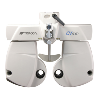
1 Hold the control lever and pull the instrument to the utmost limit toward the operator. As the
internal fixation target turns on, instruct the patient to look at the fixation target in the center.
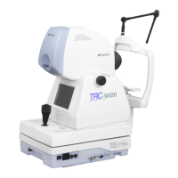
Observe the anterior segment image on the touch display.
2 Move the instrument body in right and left / up and down directions with the control lever until
you get the patient's eye in the center of the fundus live image area.
3 On the touch display, bring the ( ) scale towards the patient's pupil, and make sure that the
pupil is larger than the ( ) scale.
NOTE
Now hold the control lever upright, which will facilitate the total alignment pro-
cess.
NOTE
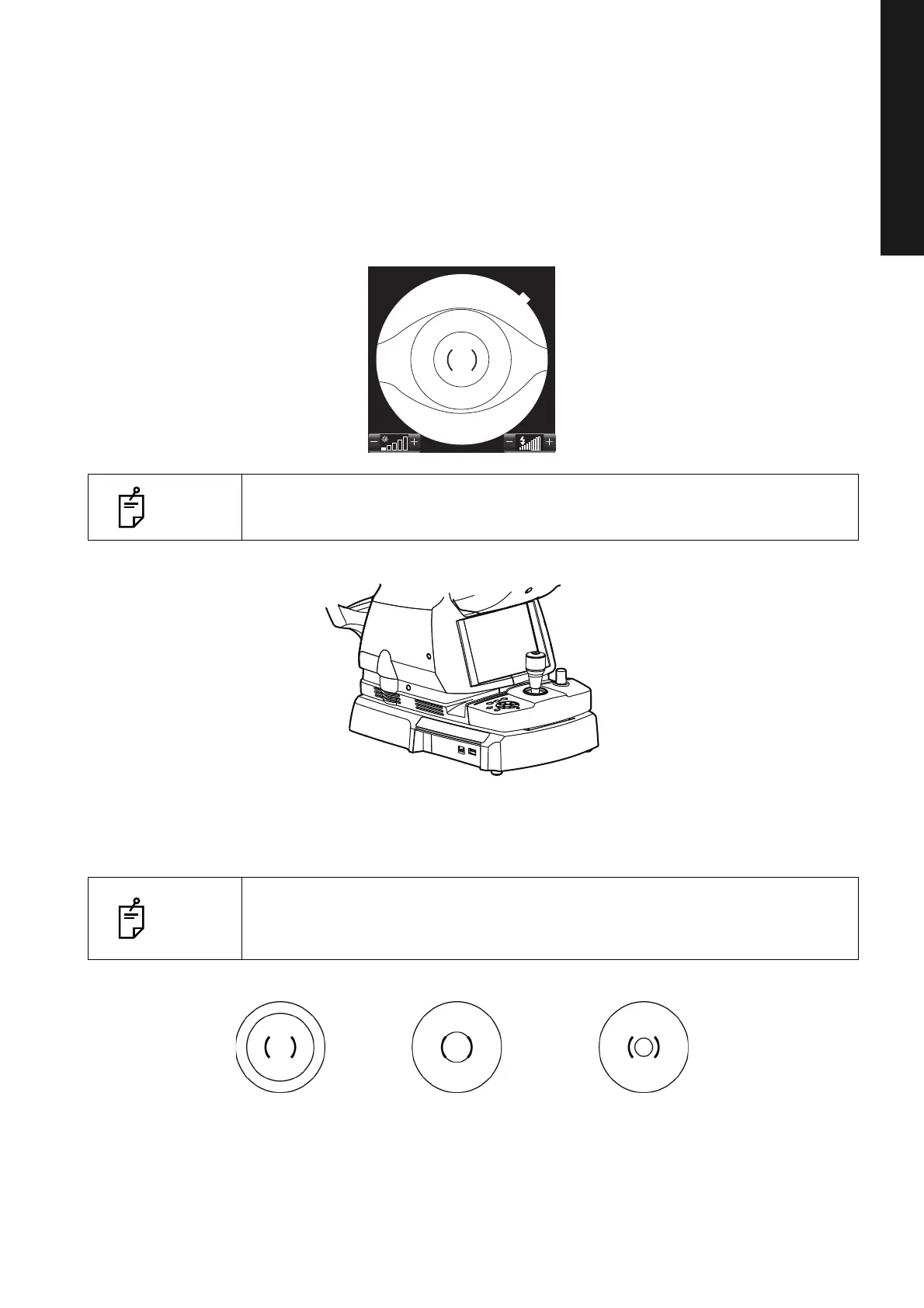
Comparison of the ( ) scale and the pupil tells you whether the pupil is large
enough for fundus photography. Use this comparison to get the standard for
photography. The diameter of the ( ) scale is approx. 4.0mm.
Well dilated. Narrowly dilated for
photography.
Pupil diameter is too small:
darken the room and further
dilate the pupil.
Pupil diameter > φ4.0mm
Pupil diameter = φ4.0mm Pupil diameter < φ4.0mm

 Loading...