70
OBJECTIVE OPERATIONS
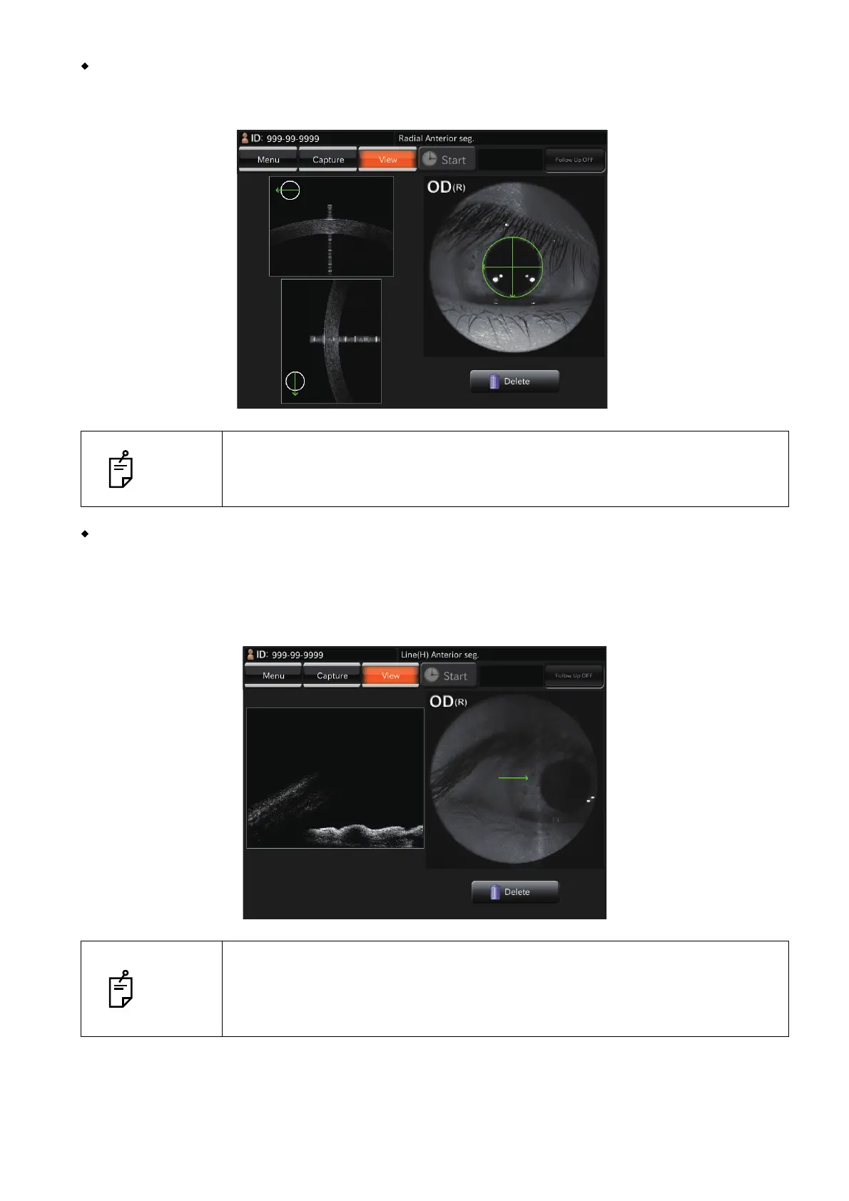
When taking a picture of cornea
Select the "Radial: Anterior segment" and move the instrument body until the cornea tomogram is displayed
near the optimal display position frame on the top of the screen.
When taking a picture of anterior chamber angle
Select "Line: Anterior segment". Push in the instrument body, which is in the pulled status. You
will see the anterior chamber angle which is upside-down. Push in the instrument body until the
tomogram of the anterior chamber angle is displayed near the optimal display position frame on
the bottom of the screen.
NOTE
Take a picture of cornea where the signal of one vertical line displayed on the
cornea tomogram is strong. This is the status where the scan center is fit to the
cornea vertex.
NOTE
When moving the tomogram of the anterior chamber angle to the lower end of
the screen, the tomogram has many folded parts. Move the instrument body to
move the anterior chamber angle display position upward a little. Then, take a
picture.

 Loading...