46
User’s Manual
CellaVision
®
DM1200
4.1.3 Estimating Platelets
The complete PLT overview image (same image as for RBC) corresponds to the
area of 8 microscopic high power fields (HPF) (100x objective and a 22 mm
ocular). The overview image is divided into 4, 9 or 16 sub-images (grid squares) as
defined by the grid size. The grid size options are 2x2, 3x3 and 4x4. The 4x4 grid
size gives the largest magnification of the image. There are as many entry fields as
there are grid squares.
Note! Help Lines are disabled for images captured on a CellaVision Image
Capture System.
There are two ways, or modes to perform the PLT estimation. This is determined in
PLT settings (see 9.6 Adjusting PLT Settings).
1. Counting PLTs in the overview image
2. Estimating the PLT concentration level
Note! When a slide is opened for the first time, the slide gets mode according to the
settings and the mode can then not be changed for that order. The system ensures
that all slides in a multi-slide order have the same mode.
Note! It is not possible to use the mode “Counting PLTs in the overview image”
for images captured on a CellaVision Image Capture System.
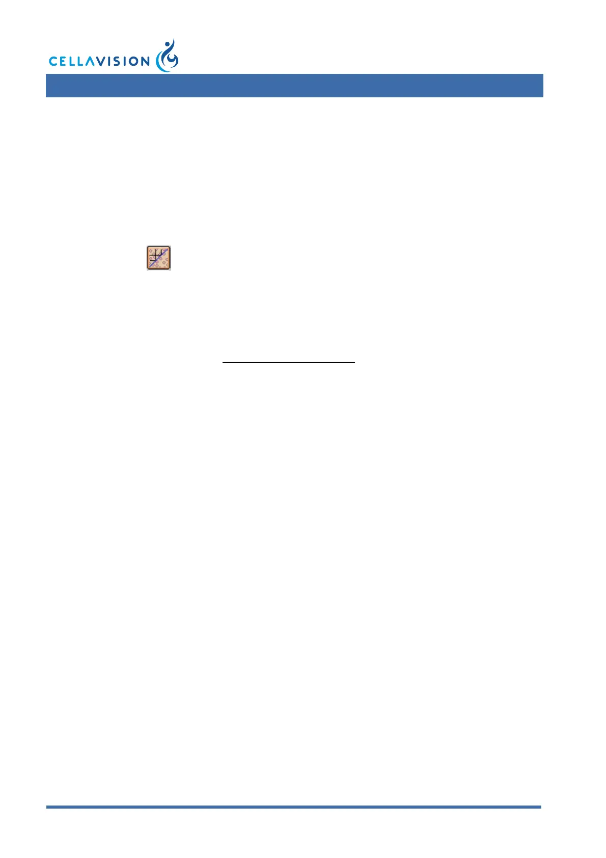
By clicking Help Lines in the toolbar, a grid of lines is drawn over the
image to facilitate the counting of PLTs.