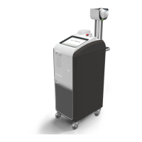
Wavelength
Skin
T
ype
Spot
Size
Window
Temperature
Treatment
Fluence
Pulse
Duration
Lesion Types
1064 nm I-V
3 mm 4-8°C 120-180 J/cm² 10-30 ms
Facial Telangiectasia,
Fine Red Spider
Veins, Cherry or
Spider Angiomas
5 mm 8°C 100-160 J/cm² 15-40 ms
Facial Telangiectasia,
Red or Blue Spider
Veins,
Periorbital Blue
Veins, Venous Lake
7 mm 8°C 110-170 J/cm² 30-60 ms
Reticular Leg Veins
2-4 mm
755 nm I-III
5 mm 4°C 50-70 J/cm² 3 ms
Facial Telangiectasia,
Superficial Blue
Vessels
8 mm 4°C 40-60 J/cm² 3 ms
Facial Telangiectasia,
Diffuse Redness
75
EXCEL HR OPERATOR MANUAL
D1796, REV. D, 12/16
Vascular Lesion Technique and Endpoints
• Ensure that the handpiece is in full contact with the skin during treatment.
- Pay particular attention when treating rounded/bony areas.
-
Precool the skin to help prevent epidermal damage.
- Ensure that each pulse receives both pre- and postcooling.
- The length of pre- and postcooling required will vary according to size, color, and
depth of vessel.
- Larger, darker vessels require longer pre- and postcooling.
• Do not stack pulses or double pulse.
- For smaller vessels, place treatment pulses adjacent or slightly spaced (avoiding
overlap).
-
For larger vessels, leave a half spot size to full spot size space between pulses.
• Endpoints will vary based on type, size, color, volume, pressure, and location of the vein.
- Common endpoints are color change, vein disappearance, and constriction.
-
If the clinical endpoint is not reached, shorten the pulse duration. If the clinical
endpoint is still not reached, then increase the fluence.
- The endpoint may not be evident or may be very subtle when treating larger
reticular leg veins.
Vascular Lesion Parameter Guidelines