ThinPrep
®
Integrated Imager Operator’s Manual
4.1
OPERATION
Chapter Four
Operation
OVERVIEW
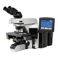
The ThinPrep
®
Integrated Imager images prepared ThinPrep Pap Test cervical cytology microscope
slides. The slides are reviewed by a cytotechnologist. The instrument may also be used as a conven
-
tional microscope, for viewing slides not associated with the ThinPrep imaging process.
Slide Preparation
Proper slide preparation is critical to successful imaging of the ThinPrep Pap Test microscope slide.
Prior to being imaged on the Integrated Imager, the slide must be:
• Processed on a ThinPrep
®
2000 System or a ThinPrep
®
5000 Processor using microscope
slides for use with the Integrated Imager (has fiducial marks)
• Stained, using ThinPrep Stain
• Coverslipped (allowed to dry thoroughly)
• Labeled in a format for use with the Integrated Imager
For the processes listed above, please consult the appropriate user documentation that came with the
equipment.
Imaging
The Integrated Imager will automatically image a slide after scanning a valid slide accession ID that
is not already in the database.
CAUTION:
Do not handle the instrument during imaging.
Proper lighting and focus of the slide is critical to successful imaging. The system disables manual
stage, focus and illumination controls. The operator should not interact with the Integrated Imager
during the approximately 90 seconds it takes to image a slide.
Slide Review
Auto Review
In this manual, Auto Review refers to a slide review in which the Integrated Imager:
• scans the slide ID number from the slide
• communicates with the database for appropriate slide data record