RETeval Flicker Option
RET
eval
Device User Manual 25
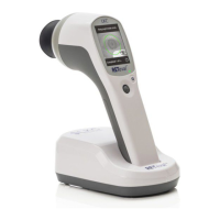
RETeval Flicker Option
The RET
eval
device measures flicker implicit time quickly and accurately by flashing light into the
patient’s eye and measuring the time delay (implicit time) and amplitude of the retina’s electrical
response as detected on the skin below the eye. The device’s patented technology enables
measurements without dilating eye drops using real-time pupil size compensation and uses skin
electrodes (Sensor Strips). The entire testing process for one patient should take less than 5
minutes.
Flicker implicit time has been correlated with a number of diseases of the retina, including
retinitis pigmentosa (Berson 1993), enhanced S-cone syndrome (Audo et al. 2008), CRVO (Miyata
et al. 2018), diabetic retinopathy (Fukuo et al. 2016, Zeng et al. 2019). Flicker implicit time has
also been used in testing preterm infants for retinopathy of prematurity (ROP) (Kennedy et al.
1997) and in identifying retinal toxicity from the anti-seizure drug vigabatrin (Miller et al. 1999,
Johnson et al. 2000, FDA Advisory Committee 2009, Ji et al. 2019). Flicker tests have been
successful in distinguishing pediatric patients with nystagmus between those with and without a
primary retinal disorder (Grace et al. 2017).
Through a protocol chooser, the test protocol can be selected from more than 10 flicker options,
including one specifically designed for vision-threatening diabetic retinopathy described earlier.
Flicker protocols
The RET
eval
device supports flicker ERG testing. Brief flashes of light are provided at the
beginning of each stimulus period. For example, the built-in protocols use a stimulus frequency
of about 28.3 Hz. Background illumination, where present, uses a PWM frequency near 1 kHz,
which is well above the human critical fusion frequency and therefore is perceived as steady
illumination.
Built-in flicker protocols typically record between 5 and 15 seconds of data for each stimulus
condition stopping after an internal precision metric is reached. Some protocols have multiple
stimulus conditions which are presented sequentially with a short (< 1 s) dark pause between the
conditions. A counter on the screen shows progress for these multi-stimulus protocols.
Many of the protocols have constant retinal illuminance, which are described by the Troland unit
(Td). These protocols are identified with “Td” in the user interface and PDF reports. In these
protocols, the RET
eval
device measures the pupil size in real time and continuously adjusts the
flash luminance to deliver the desired amount of light into the eye regardless of the size of the
pupil according to the following formula: Troland = (pupil area in mm
2
)(luminance in cd/m
2
).
Thus, pupils do not need to be dilated to achieve consistent results. Even when using mydriatics,
people dilate to different diameters and results can be made more consistent by using the
Troland-based stimuli. While Troland-based tests make results less dependent on pupil size,
secondary factors such as the Stiles-Crawford effect and/or changes in the distribution of light
on the retina prevent Troland-based tests from being completely independent of pupil size (Kato
et al. 2015, Davis, Kraszewska, and Manning 2017, Sugawara et al. 2020).
Stimuli having flash retinal illuminance energies of 4, 8, 16, and 32 Td∙s of white light (1931 CIE x,
y of 0.33, 0.33) without background illumination are provided.