Chapter 2 - Introduction
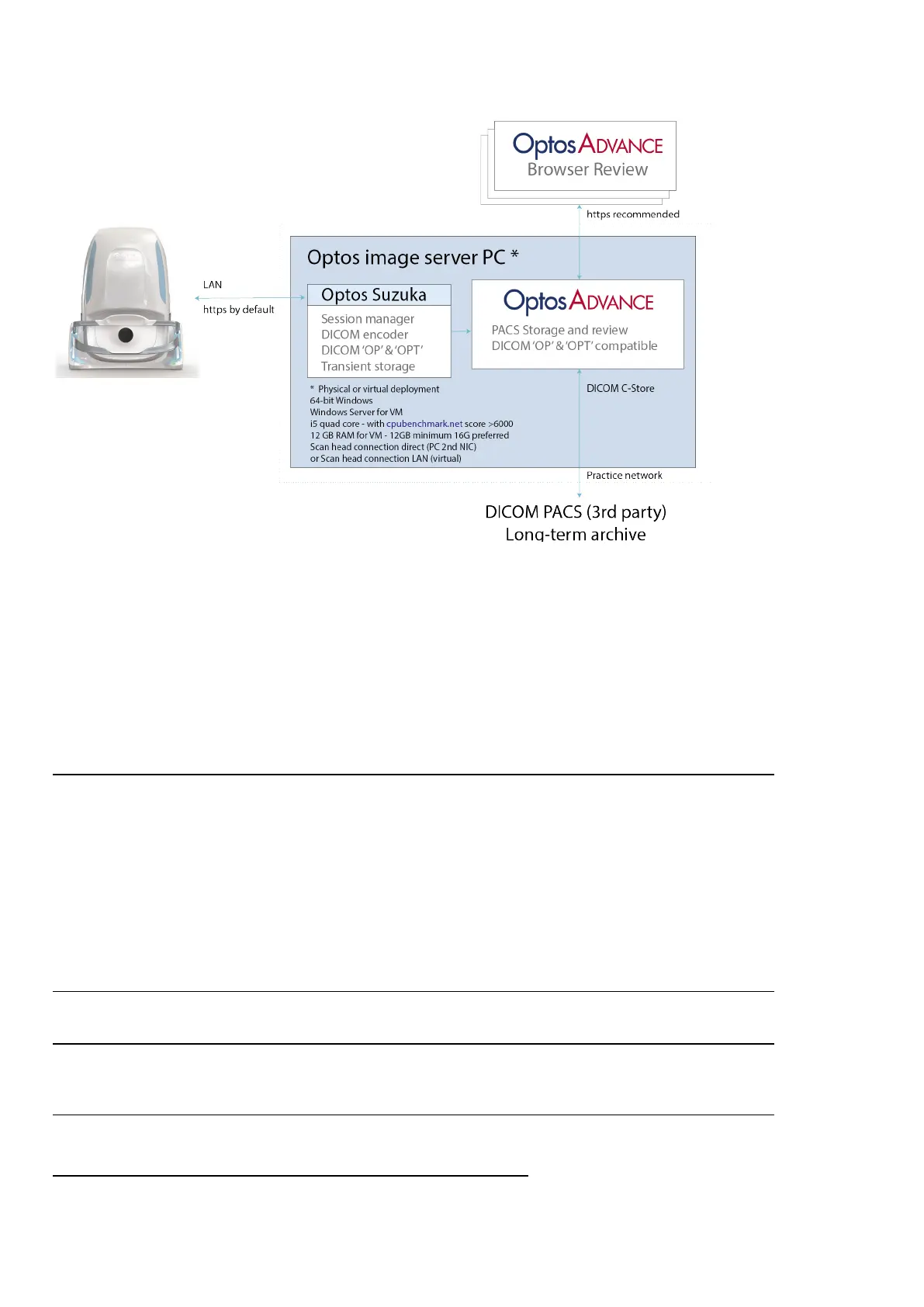
FIGURE 2: Network data flow
2.6.3 Image modalities
Optos multi-modal ultra-widefield retinal imaging enables the capture of posterior images, with a field of view
of up to 200 degrees in a single capture, and in an imaging time of less than 0.4 seconds.
Depending on the configuration of your device the following image modalities may be available:
2.6.3.1 SLO imaging modalities
optomap
optomap is an ultra-widefield (UWF
1
) color fundus image.
optomap combines neurosensory retinal and choroidal reflectance images and provides UWF image views
from a single capture action:
l optomap color view – an UWF color fundus image combining red and green reflectance images.
l optomap neurosensory retinal view – an UWF green laser reflectance image of the surface retina.
l optomap choroidal view – an UWF red laser reflectance image of the deeper retina.
optomap af
optomap af is an UWF green laser auto fluorescence image.
optomap fa
optomap fa is an UWF fluorescein angiography (FA) image.
optomap icg
optomap icg is an UWF indocyanine green (ICG) angiography image.
1.
Ultra-widefield images provide a field of view of up to 200° in a single capture.
Page 24 of 72 Part Number: G110230/1ENG
English Copyright 2019, Optos plc. All rights reserved.