Chapter 2 - Introduction
Optos OCT retina scan
Captures a high density raster scan of OCT images covering a 30 x 30 degrees (9 mm x 9 mm) field of view
and centered around the macula. The Optos OCT retina scan comprises 111 horizontal line scans.
FIGURE 5: Optos OCT retina scan example scan area and cross hairs
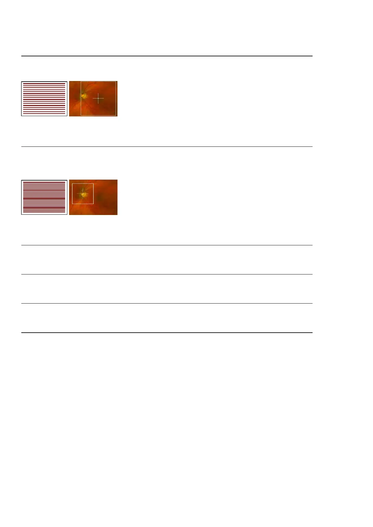
Optos OCT optic nerve head scan
Captures a high density raster scan of OCTimagescovering a 20 x 20 degrees (6 mm x 6 mm) field of view
centered around the optic nerve head. The Optos OCT optic nerve head scan comprises 111 horizontal line
scans.
FIGURE 6: Optos OCT optic nerve head scan example target and scan lines
Optos OCT ultra-widefield line scan
The ultra-widefield line scan can be positioned anywhere within an on-axis optomap image. It generates a
cross-sectional OCT image of 20 degrees (6 mm) in length.
Optos OCT ultra-widefield volume scan
Volume scans can be positioned anywhere within an on-axis optomap image. The scan captures 121 B-
scans covering a 20 x 20 degrees (6 mm x 6 mm) field of view.
Optos OCT ultra-widefield HD volume scan
HD volume scans can be positioned anywhere within an on-axis optomap image. The high density scan
captures 121 B-scans covering a 20 x 12 degrees (6 mm x 3 mm) field of view.
Optos OCT ultra-widefield extended line scan
The extended line scan can be positioned centrally within an on-axis optomap image. It generates a cross-
sectional OCT image of 80 degrees (23 mm) in length.
2.6.3.3 Multi-mode workflow
The device provides a multi-modal workflow sequencing option. Multi-mode assists the user in performing a
sequence of SLO and OCT scans, see Capture using Multi-mode on page47.
2.6.4 System software
The system contains features to help you configure the system and to capture, review, archive and retrieve
images. The system software comprises:
l Scan head software that enables capture of patient studies.
l Image server 'middleware' that encodes patient studies for storage.
l Image server 'archive and review' software provided by OptosAdvance.
l Browser-based review clients.
The image capture software runs on the scan head.
Page 26 of 72 Part Number: G110230/1ENG
English Copyright 2019, Optos plc. All rights reserved.