3-29
The following items are used for checking to see if ultra-
sound can correctly catch the axial length and, therefore,
reasonable waveforms are obtainable, provided that these
items are not good conditions for taking-in of measurement
data.
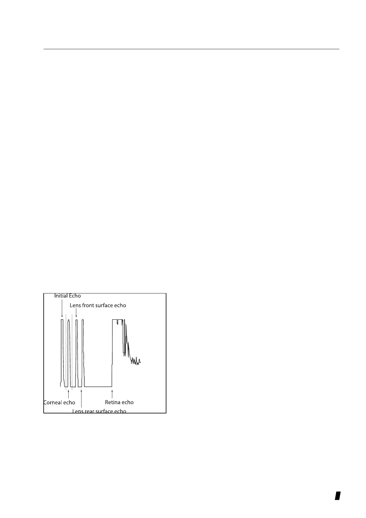
1) The retinal echoes rise high.: implies that the probe is
applied perpendicular to the cornea.
2) The echo rises high in the front and at the rear of the
crystalline lens: implies that the axial length was caught
with the probe.
3) The retinal and sclera echoes are distinctive.: implies
that the probe is applied at a right angle. In case of high
gain, the drop (choroid) between two echoes cannot be
identified, which is not always necessary to be detected.
4) No tail waves followed after corneal echos.: implies that
the probe directly touches the cornea. If there is any tear
or corneal protective gel left between the cornea and the
probe, the corneal echoes are followed by tail waves. If
so, the measurement of axial length may not be stable or
may be longer than its actual length.
b) Immersion mode
For immersion mode, the following conditions are added
to those of Contact mode.
The cornea front echoes must be within the range of 1.8
and 3.2 mm (which is shown as the range of dotted line in
the left figure.)
The following items are not to indicate the conditions of
data acquisition, but to confirm if acquired waveform is
acceptable or not.
Also confirm “a Contact mode i - iii ”.
1) No unnecessary echoes arise if there is air voids
included in the ultrasound gel used in the inside of the
tip of the immersion attachment or between the probe
and the cornea.
 Loading...