136
User’s Manual
CellaVision
®
DM1200
(Cont'd)
Performance Specification Peripheral blood
Average WBC cell-location and display of at least 97 % with a standard deviation
less than 2 %.
Throughput*: Approximately 20 slides/h for complete orders containing RBC,
PLT and 100-cell WBC.
Results of short-term imprecision found in a clinical evaluation on 230 patient
samples, based on NCCLS standard H-20A:
Limitations: Distinctions between band and segmented neutrophils,
metamyelocytes and myelocytes, myelocytes and promyelocytes, lymphocyte and
lymphocytes variant forms are subjects to variations among individual operators.
Performance Specification Body Fluid
Average WBC cell-location and display of at least 97 % with a standard deviation
less than 2%.
Throughput*: Approximately 15 slides/h for orders (100 WBCs) containing only
10x overview images (6 mm analysis area). Approximately 3 slides/h for orders
(100 WBCs) containing both 10x and 50x overview images (6 mm analysis area).
To evaluate the Accuracy and Short-term imprecision of the Body Fluid
application for DM1200, a comparison study was conducted at two sites with 5
operators. CellaVision DM1200 running the body fluid application was compared
to CellaVision DM96 running the body fluid application. 200-cell differential
counts were performed by qualified blood examiners. The study was performed
according to CLSI EP9A-2 Method Comparison and Bias Estimation Using Patient
Samples; Approved Guideline. The short-term imprecision was calculated as
defined in CLSI H56-A Body Fluid Analysis for Cellular Composition; Approved
Guideline. The results are presented in the tables on the next page.
* “Depending on WBC concentration, number of non-WBCs and the quality of the
smear.”
(Cont'd)
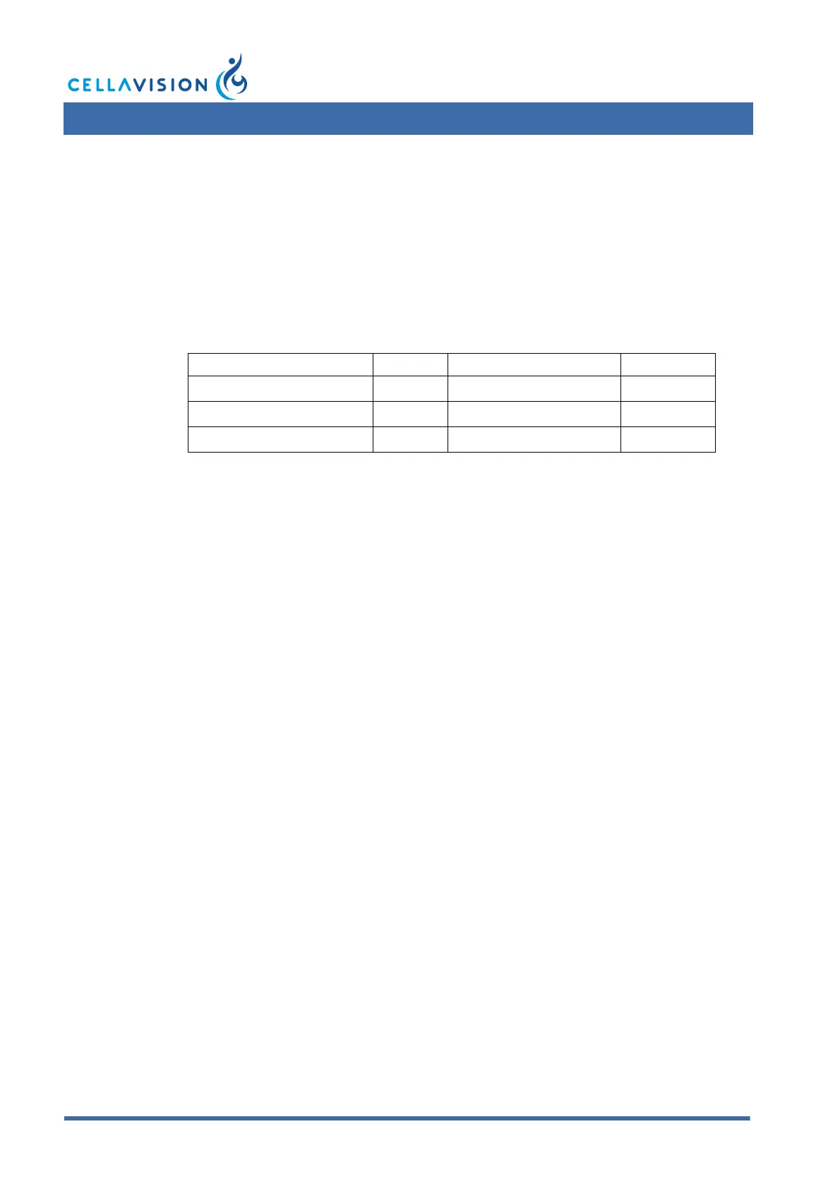
Cell class SD (%) Cell class SD (%)
Segmented neutrophils 3.8 Basophils 0.7
Band neutrophils 1.6 Lymphocytes 3.4
Eosinophils 1.0 Monocytes 2.0
 Loading...