om 5184516-100 Rev. 5 6-26
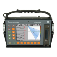
4. Select the Cursor Function pull–down list shown in bottom, right
corner of the
image portion of the screen.
5. Select the Stenosis option. The feature menu on the left will change to show
the Stenosis feature buttons.
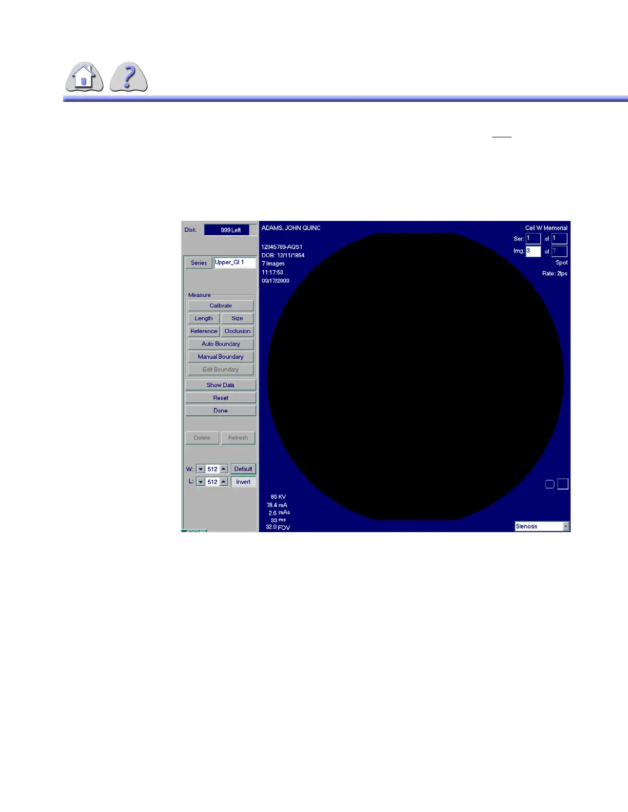
ILLUSTRATION 6-18
STENOSIS SCREEN
18-1Calibration
Calibrating the system involves the operator defining the outer boundaries of a
known diameter. It is important that the calibration be performed accurately, since
this procedure allows the system to assign a millimeter value to the “width” of
each pixel of the image. This allows for accurate measurements.
The following size Image Intensifier and catheter size combinations are NOT per-
mitted:
12” II (32 cm) 3F
16” II (40 cm) 3F & 4F
FOR TRAINING PURPOSES ONLY!
NOTE: Once downloaded, this document is UNCONTROLLED, and therefore may not be the latest revision. Always confirm revision status against a validated source (ie CDL).