Do you have a question about the GE Precision 500D and is the answer not in the manual?
Provides necessary information for proper system operation, not for clinical diagnosis.
Requires sufficient knowledge to perform diagnostics imaging procedures.
Describes symbols for danger, warning, and caution used for safety instructions.
Indicates that electrical and electronic equipment waste must be disposed of separately.
Explains safety considerations, equipment/patient precautions, and symbols for safe operation.
Provides information and precautionary measures for safe system operation.
Details safety considerations for X-ray equipment operation and potential hazards.
Describes measurements of stray radiation distribution around the GE Precision 500D R & F unit.
Outlines general precautions for safe operation and hazard awareness.
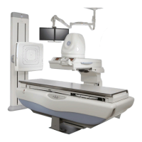
Introduces the P500D as a high-resolution, PC-based digital imaging system.
Details operating, transport, and storage environmental conditions.
Explains the system's capability for storing various types of images.
Describes optional features such as DICOM 3.0 Interface and Bar Code Scanner.
Provides instructions for powering the system on and off safely.
Describes the graphical user interface and input methods.
Explains how to select X-ray tube 1 for fluorography and tube 2 for radiography.
Explains system startup, shutdown, and tube warmup procedures.
Details the concepts for performing routine startup and shutdown procedures.
Explains the functionality of the integrated user interface.
Describes the scenario for starting an exam by selecting a patient.
Provides tools for creating, modifying, and deleting protocol categories and protocols.
Explains the digital image review screen and utility menu.
Allows changing the series name for descriptive purposes.
Displays patient information and details functionality of the patient list screen.
Explains the optional feature to send images to a reference monitor.
Describes how to select a patient file for review on the digital image monitor.
Provides instructions for acquiring images and performing Quality Assurance tests.
Details the feature to consolidate a series of images into a single image.
Explains how to adjust image contrast and window settings.
Explains how to adjust image brightness and level settings.
Describes the function of the invert button for image polarity.
Explains how to set default image parameters.
Details the zoom feature for magnifying images.
Explains the system's capability to apply edge enhancement for spatial resolution.
Describes optimizing images using features like Window, Level, Invert, Edge, Zoom.
Explains how to view multiple images from a patient file or series.
Provides instructions for deleting images during acquisition or from the hard drive.
Explains how to add, edit, and move text annotations and pointers on images.
States that all printing is done via the IUI console, referring to Chapter 5.
Details features and steps for performing stenosis measurements.
Explains how to search DICOM Worklist databases for patient information.
Introduces the Intelligent Digital Device (IDD) as an image receptor.
Details system identification and compliance plates as shown in illustrations and tables.
Describes the X-ray exposure modes: Digital Record and Fluoro.
Illustrates travel specifications for the Intelligent Digital Device and table top.
Explains that control of digital operation is from the IDD film control panel.
Lists types of radiological examinations for which the Significant Zone of Occupancy is designated.
Locates operator controls and displays in two basic areas: Table Side and Imaging Device.
Provides an overview of imaging device operator controls and displays.
Details basic controls and displays on the main operator console.
Describes optional controls and displays on the main operator console.
Details the controls and displays on the auxiliary digital operator console.
Describes the controls for digital fluoro presentation.
Details the controls for the digital multi-function operator control group.
Illustrates and describes operator controls and displays for the Precision 500D table.
Provides product specifications for the Precision 500D Table.
Details specifications for tables, tubes, and collimators.
Covers operational procedures for the imaging device carriage.
Explains how to perform fluoro exposures using buttons or footswitch.
Details using the Prep/Record Bar and Integrated Fluoro/Expose Switch.
Provides instructions for equipment positioning for table bucky procedures.
Introduces the Overhead Tube Suspension (OTS) as a positioning device.
Details system identification and compliance labels.
Describes the overhead rail system components and operation.
Explains the function of the telescopic column and carriage.
Details the use of longitudinal and transverse detents for positioning.
Explains how to rotate the tube unit about the vertical axis.
Describes tube angulation about the short axis and its controls.
Explains the in-room user interface for receptor type, kV, and mAs selections.
Details the controls for the automatic collimator.
Explains the function of the locking lever for securing accessories.
Describes the automatic collimator display and its indicators.
Illustrates the bottom view of the automatic collimator.
Explains the linear laser light localizer for longitudinal centering.
Describes the centering cross for displaying longitudinal and transverse axes.
Shows the rear view of the collimator, including the locking lever.
Provides instructions for changing collimator lamps.
Provides instructions for changing lamps on Model 5234954 collimator.
Explains how to rotate the collimator 90° around its vertical axis.
Defines the SG80/120 as a Vertical Bucky Stand for radiographic examinations.
Details the main parts of the SG80/120, including Column Assembly.
Describes applications for SG80/120 vertical Bucky stands.
Covers operational procedures for vertical positioning of the Bucky.
Explains how the Bucky is held and moved vertically.
Details how to load a cassette into the bucky assembly tray.
Provides procedure for removing a cassette from the manual tray.
Explains AEC detector areas and corresponds to front panel patterns.
Emphasizes X-ray tube centering with the Bucky for accurate alignment.
Describes accessing the bucky mechanism and grid by removing the front panel.
Details SG120-specific functionalities like Bucky Rotation.
Outlines the importance of a periodic maintenance program for safety.
Shows required labels for SG80 and SG120 units.
Lists electrical requirements for the SG80/120 vertical bucky stand.
Provides dimensions for the SG80, SG80 W/SPACER, and SG120.
Specifies crate dimensions for the SG80/120.
Details general and bucky specifications for the SG120.
Defines the SG-100/SG-120 as a Tilting/Rotating Vertical Bucky Stand.
Illustrates and labels the components of the SG-100.
Provides essential precautions for safe equipment operation.
Details general description and vertical positioning of the Bucky.
Explains how the Bucky is held and moved vertically.
Describes the use and safety of patient support legs when Bucky is horizontal.
Explains how to rotate the Bucky from 0° to 180° using the lock lever.
Details the angulation range and control of the SG120 bucky assembly.
Describes how to open the hinged front panel for access.
Details how the SG-100 is equipped with a manual cassette tray.
Explains how the manual cassette tray accepts and holds cassettes.
Provides procedure for removing a cassette from the manual tray.
Explains AEC detector areas and corresponds to front panel patterns.
Emphasizes X-ray tube centering with the Bucky for accurate alignment.
Explains requirements for accurate positioning for automatic collimation.
Provides general dimensions and weight for the SG-100.
Details the dimensions of the column assembly.
Provides dimensions and filtration for the front panel.
Lists power requirements for the unit.
Shows general dimensions for the bucky.
Lists available accessories and options for the SG-100.
Introduces the JEDI generator models and specifications.
Discusses precautions and site modifications for radiation protection.
Provides specifications for the JEDI generator system cabinets.
Details general cabinet specifications.
Lists model-dependent specifications for input power.
Details generator output power specifications.
Specifies minimum wire size requirements for 65 KW generator.
Explains X-ray control safety switch and tube housing overheat interlock.
Details table accessories, including patient step and foot rest.
Lists optional table accessories like shoulder rest and compression device.
Explains setup for myelographic procedures, including horizontal stop.
Describes optional myelographic boots for patient support.
Details the installation and use of the optional arm board.
Describes the attachment and adjustment of optional knee crutches.
Explains how to install the vertical cassette holder for cross-table radiography.
Describes the Maxiray 100 as a high-speed tube unit for radiography.
Highlights features like Polyrhenium target and anode heat storage capacity.
Details tube housing construction and environmental specifications.
Describes the glass envelope and anode/cathode design for heat dissipation.
Provides heat storage capacity and maximum heat dissipation rates.
Explains tube rating charts based on target temperature.
Details focal spot sizes and thermal limitations for fluoroscopy.
States that published Company warranty applies.
Outlines routine cleaning and major maintenance by qualified personnel.
Lists UL and CSA certifications and product standards.
Describes the first exposures of the day for system and tube function testing.
Emphasizes the need for a periodic maintenance program for safe performance.
Outlines the owner's responsibility for supplying or arranging periodic maintenance.
Highlights the need for service personnel trained on medical x-ray apparatus.
Advises caution with equipment safeguards and proper cleaning procedures.
Contains instructions on how to perform the Quality Assurance Process (QAP).
Provides step-by-step instructions for performing the QAP.
Defines operating modes: Fluoro, Spot Film, Digital Record, Table Bucky.
Defines operating modes: Fluoro, Spot Film, Digital Record, Table Bucky.
Defines terms like SID, SOD, and Field of View (FOV).
Explains symbols used on system hardware.
Explains symbols used on system hardware.
Describes the Fluoroscopy imaging technique and its use.
Explains how to engage Fluoroscopy mode by selecting 'Digital'.
Lists types of radiological examinations for Fluoroscopy mode.
Specifies mode usage is directed by a physician specializing in diagnostic imaging.
Describes Pulsed Fluoroscopy as a dose reduction technique.
Explains how to engage Pulsed Fluoroscopy mode by selecting 'Dig Pulsed'.
Describes DSA as a purchasable option for visualizing blood vessels.
Explains DSA mode engagement via IUI interface screen selection.
Describes standard X-ray exam using a cassette.
Explains engaging Radiographic Mode via 'Start Cassette Only Exam'.
| Brand | GE |
|---|---|
| Model | Precision 500D |
| Category | Measuring Instruments |
| Language | English |