om 5184516-100 Rev. 5 2-6
For a system, the location of the CE marking label is described in the system
manual.
European registered place of business:
GE Medical Systems Europe
Quality Assurance Manager
BP 34F 78533 BUC CEDEX
France
Tel: +33 1 30 70 40 40
• Green QSD 1990 Standard issued by MDD (Medical Devices Directorate,
Department of Health, UK).
• Medical Device Good Manufacturing Practice Manual issued by the FDA
(Food and Drug Administration, Department of Health, USA).
• Underwriters’ Laboratories, Inc. (UL), an independent testing laboratory.
• Canadian Standards Association (CSA).
• International Electrotechnical Commission (IEC). International standards
organization, when applicable.
General Electric Medical Systems is ISO 9001 certified.
SECTION 4
RADIATION SURVEY
4-1Introduction
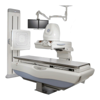
This report describes measurements of the distribution of stray radiation around
the GE Precision 500D R & F unit. These measurements were made as specified
in IEC 60601.
4-2Materials and Methods
The reference point for all measurements was chosen as the center of the table-
top with the table positioned horizontally, using its centering function. All measure-
ments were made with a phantom consisting of a 25 cm cube of polymethyl
methacrylate (PMMA) centered on the tabletop. The entrance surface of the
image intensifier was set at a distance of 30 cm above the tabletop. The nominal
field size was 22 cm. The x–ray tube voltage was at the maximum fluoroscopic
value of 120 kVp and the x–ray tube current was 4.6 mA. This resulted in an
entrance area kerma product of 900 RVcm/min. This was used to normalize all of
the measured values of stray radiation.
The unit was placed in four typical configurations for the surveys. Two of the con-
figurations had the tabletop horizontal and the x–ray beam vertical and centered
to the table. In this position, measurements were made without and with the pro-
tective lead curtain attached to the image intensifier tower. In these configura-
tions, the central ray of the x–ray beam corresponded to the vertical projection of
the chosen reference point. A third configuration was with the table vertical and
the x–ray beam horizontal. The focal spot of the x–ray beam in this case was 125
cm above the floor. The protective lead curtain was not attached to the image
intensifier tower, but the footrest was fitted to the table in this configuration. The
FOR TRAINING PURPOSES ONLY!
NOTE: Once downloaded, this document is UNCONTROLLED, and therefore may not be the latest revision. Always confirm revision status against a validated source (ie CDL).