om 5184516-100 Rev. 5 D-6
3-5 Example of specific clinical procedures
Primarily used for contrast filled vessels.
DSA, an electronic technique for imaging blood vessels, is useful in diagnosing
arterial occlusion, including carotid artery stenosis and pulmonary artery thrombo-
sis, and in detecting renal vascular disease. After contrast material is injected into
an artery or vein, a physician produces images that show the difference between
the contrast-filled vessels and surrounding anatomy through an image subtraction
process.
3-6 How mode should be used
Used by or under the direction of a physician specializing in diagnostic imaging.
SECTION 4
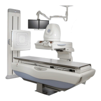
RADIOGRAPHIC MODE
4-1 Description
Standard x-ray exam using a cassette.
4-2 System Controlled Technique Factors
• Fixed or Automatically Selected Technique Factors: Fixed technique parame-
ters include: kV, mA, ms, focal spot size. In the automatic exposure control
(AEC) mode, the exposure time is determined when a pre-set density has
been reached during exposure.
• How automatic adjustment is controlled: A calibrated ion chamber controls the
duration of the exposure time.
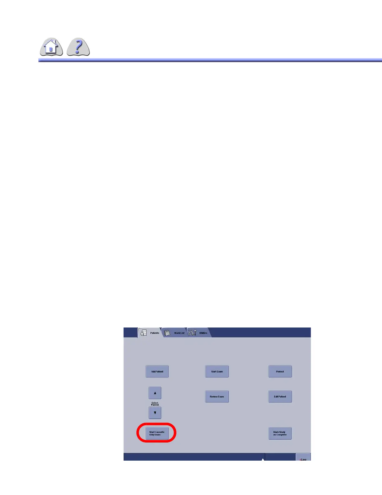
4-3 How to engage/ disengage
The mode is engaged by pressing the “Start Cassette Only Exam” button on the
IUI interface screen.
FOR TRAINING PURPOSES ONLY!
NOTE: Once downloaded, this document is UNCONTROLLED, and therefore may not be the latest revision. Always confirm revision status against a validated source (ie CDL).