MAN-05359-001 -001 Rev. 001 page 4 of 32
Review in a separate multi-center clinical study. Both clinical studies are described in the following
sections.
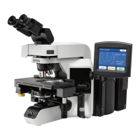
G.1 ThinPrep Imaging System Compared to Manual Review
A multi-center, two-armed clinical study was performed over an eleven (11) month period at four
(4) cytology laboratory sites within the United States
2
. The objective of the study entitled “Multi-
Center Trial Evaluating the Primary Screening Capability of the ThinPrep
®
Imaging System” was to
show that routine screening of ThinPrep Pap Test slides using the ThinPrep Imaging System is
equivalent to a manual review of ThinPrep slides for all categories used for cytologic diagnosis
(specimen adequacy and descriptive diagnosis) as defined by the Bethesda System criteria
1
.
The two-arm study approach allowed for a comparison of the cytologic interpretation (descriptive
diagnosis and specimen adequacy) from a single ThinPrep-prepared slide, screened first using
standard laboratory cervical cytology practices (Manual Review) and then after a 48-day time lag
were screened with the assistance of the ThinPrep Imaging System (Imager Review). A subset of
slides from the study were reviewed and adjudicated by a panel of three (3) independent
cytopathologists to determine a consensus diagnosis. The consensus diagnosis was used as a “gold
standard” for truth to evaluate the results of the study.
G.1.1 Laboratory and Patient Characteristics
Of the 10,359 subjects in the study, 9,550 met the requirements for inclusion in the descriptive
diagnosis analysis. During the study, 7.1% (732/10,359) slides could not be read on the Imager
and required a manual review during the Imager Review arm. Excessive number of air bubbles
on the slides was the leading contributor. Additional factors included focus problems, slide
density, slide identification read failures, slides detected out of position, multiple slides seated
within a cassette slot and slides that had already been imaged. The cytology laboratories
participating in the study were comprised of four centers. All sites selected had extensive
experience in the processing and evaluation of gynecologic ThinPrep slides, and were trained in
the use of the ThinPrep Imaging System. The study population represented diverse geographic
regions and subject populations of women who would undergo cervical screening with the
ThinPrep Imaging System in normal clinical use. These sites included both women being
routinely screened (screening population) and patients with a recent previous cervical
abnormality (referral population). The characteristics of the study sites are summarized in
Table 1.