52 Chapter 7 Acquiring a 3D Model for Orthodontics
The example below shows a 3D model of the upper jaw when the occlusal, lingual, and buccal
surfaces have been completely scanned, as well as much of the palate.
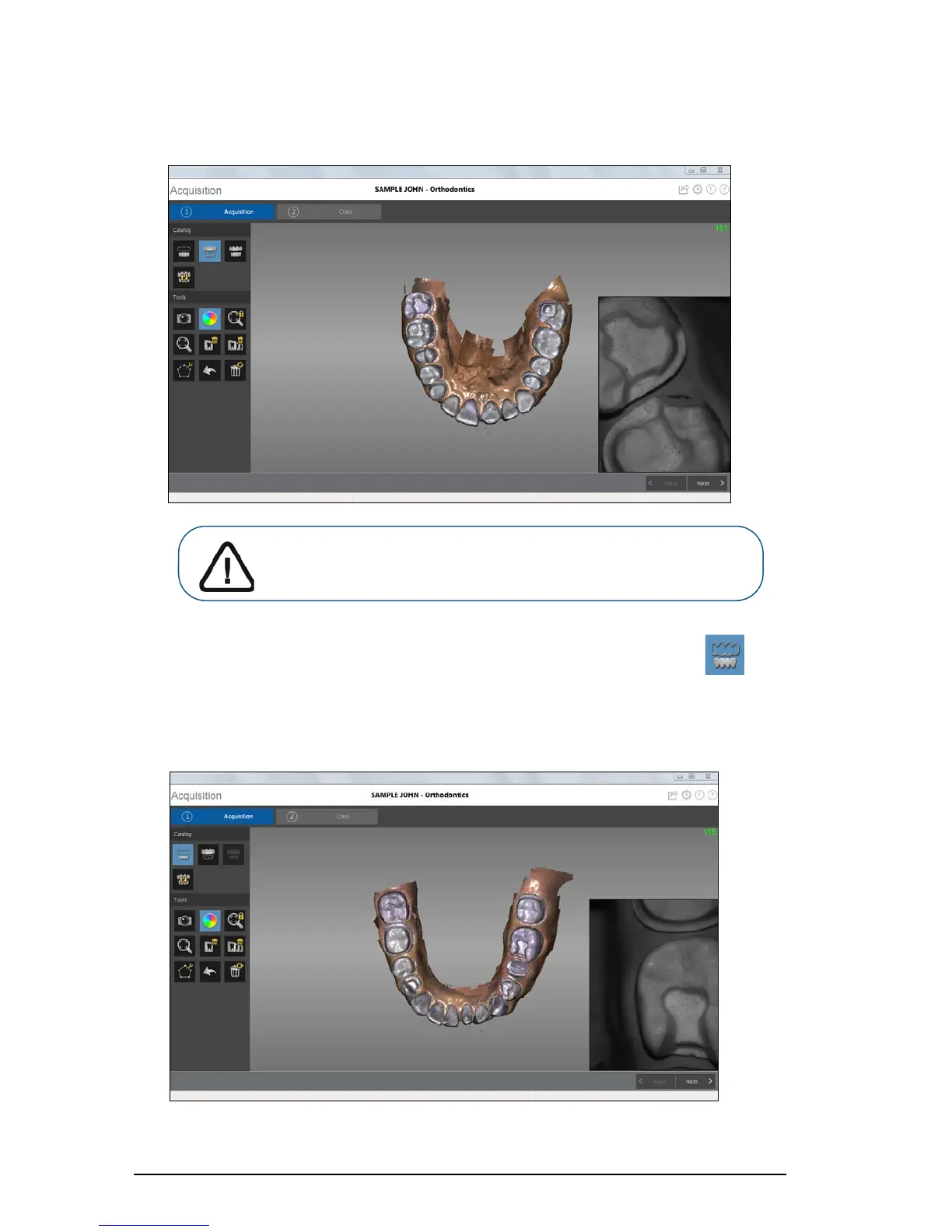
9 Once the upper jaw has been scanned, you can begin scanning the lower jaw. Click and
repeat steps 5 through 8 until the teeth on the lower jaw are scanned.
The example below shows a 3D model of the lower jaw when the occlusal, lingual, and buccal
surfaces have been completely scanned, including soft tissue.
Important: If holes are displayed in the scanned image, re-scan the
area until the holes are filled. Use the mouse wheel to zoom in on
the image for a closer look.