Do you have a question about the Planmeca Viso and is the answer not in the manual?
Details specific features and instructions for safer pediatric use.
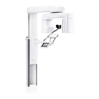
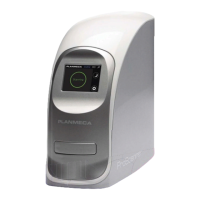
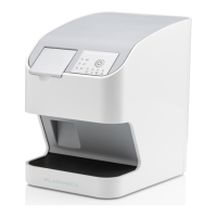
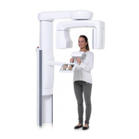
Illustrates and labels the main components of the entire X-ray system.
Detailed illustration and labeling of the X-ray unit's individual components.
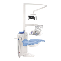
Illustrates and lists the various patient support components used for positioning.
Describes the exposure switch, its location, and indicator lights.
Explains the function and operation of the emergency stop button.
Details the touch screen and virtual control panel for operating the unit.
Steps to prepare the X-ray system, including attaching patient supports.
Detailed instructions for attaching various patient support components.
Instructions for preparing the patient, including removing accessories and applying lead apron.
Overview of available 2D programs: Panoramic, Bitewing, and 2D View.
Steps to select the desired 2D imaging program from the interface.
Detailed steps for correctly positioning the patient for 2D exposures.
How to adjust kV and mAs values for optimal 2D exposure settings.
Detailed steps for correctly positioning the patient for 3D exposures.
How to adjust the volume position and size using the on-screen interface.
How to select image resolution, ULD, and the ProFace feature for 3D imaging.
How to adjust exposure values for current 3D exposures.
Step-by-step guide for performing a 3D exposure.
Step-by-step guide for running the 3D Quality Assurance test.
| Software | Planmeca Romexis |
|---|---|
| X-ray tube voltage | 60-120 kV |
| Focal Spot Size | 0.5 mm |
| Category | Dental equipment |
| Imaging Modes | CBCT, Panoramic, Cephalometric |
| Patient Positioning | Standing, sitting |
| Imaging Modalities | CBCT, Panoramic, Cephalometric |
| X-ray tube current | 1 mA - 10 mA |
| Exposure Time Range | Varies depending on imaging mode and settings |
| Image Receptor Size | Varies depending on mode |
| Power Supply | 230 V AC |
| Sensor Type | Flat panel sensor |