Chapter 6. Viability Methods
8004708 Cellometer™ Ascend User Manual Rev A 26
Using AO/PI Viability Method
Dual-fluorescence methods have been developed to accurately determine nucleated cell concentration and
viability in primary cell samples containing debris and non-nucleated cells, including platelets and red blood cells.
In the AO/PI Viability Method, acridine orange (AO) enters all cells and stains their DNA causing nucleated cells to
fluoresce Green (470/534 Channel), while propidium iodide (PI) only enters dead cells with compromised
membranes and stains their DNA causing them to fluoresce Red (531/655 Channel).
• Cells stained with both AO and PI fluoresce Red due to quenching.
• Live nucleated cells are easily identified in the Green fluorescence channel.
• Dead nucleated cells are easily identified in the Red fluorescence channel.
As a result, debris and non-nucleated cells do not interfere with nucleated cell counts when using the AO/PI
viability method.
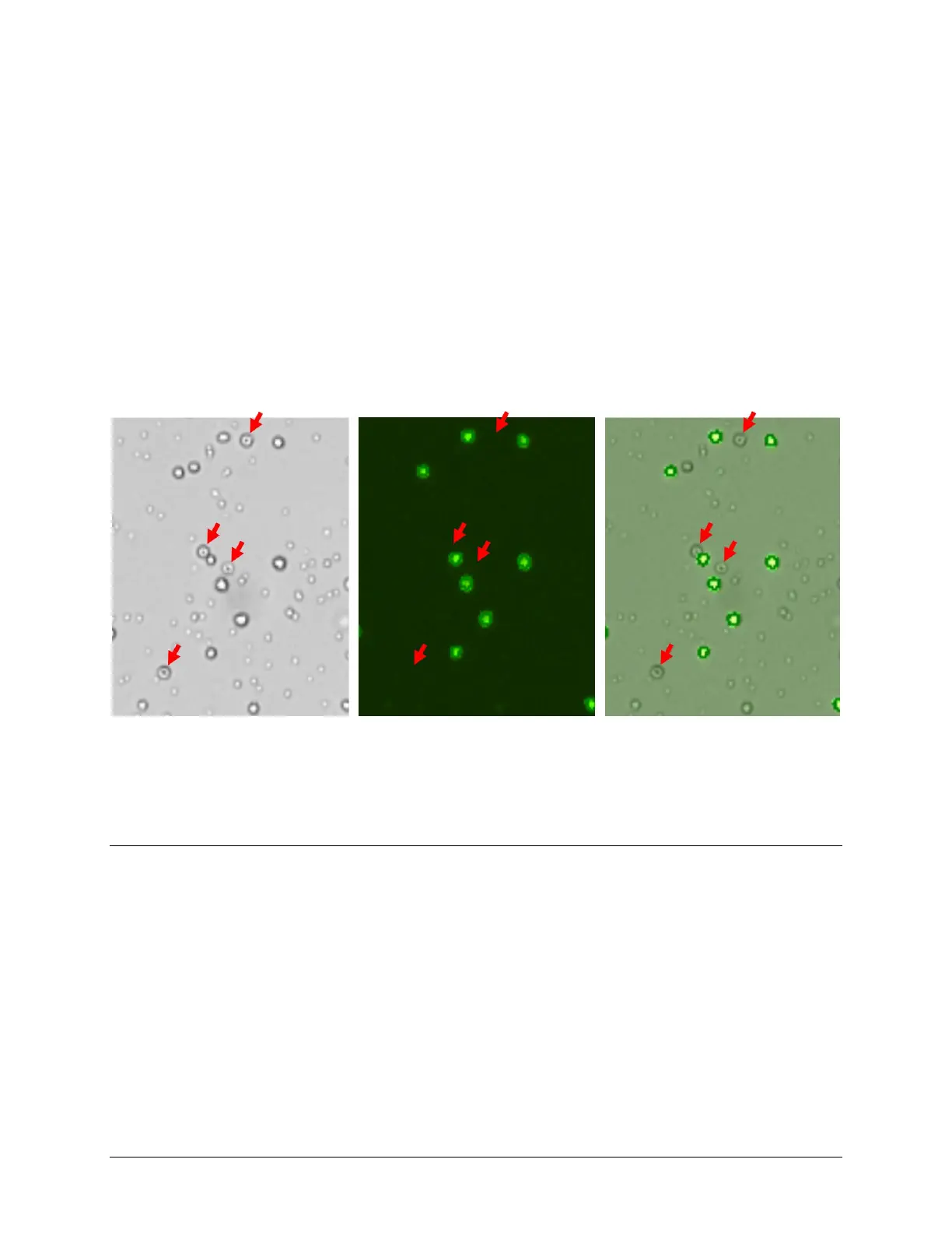
In images captured by Cellometer Ascend using the AO/PI viability method, red blood cells (RBCs, marked by red arrows)
seen in the brightfield image (on left) are not seen in the fluorescent image (in middle), but are clearly identified in the
overlay image (on right). Only nucleated cells are counted using the AO/PI staining method resulting in a more accurate total
cell count and percent viability calculation.
PREPARING A CELL SAMPLE FOR AO/PI VIABILITY DETERMINATION
Invert the tube containing cells ten times (10x) and pipette up and down 10x to generate a homogeneous cell
sample and reduce cell clumps. Do not shake or vortex the sample as this may damage cell membranes.
For viability measurement, stain cells by combining 20 μL of sample with 20 μL of AO/PI staining solution. For whole
blood and other viscous samples, draw sample in and out of the pipette tip at least once prior to transferring for
staining. Gently mix stained cell solution by pipetting up and down 10x before adding sample to counting chamber.