24 300-002547-00 rev7
Recommended CT Scan and Reconstruction Parameters
In order to produce CT images suitable for use with the Monarch Pre-Op
Planning Application and the Monarch Navigation Application, the following
conditions should be met while scanning the patient:
Patient position: Supine, immobile, full inspiration breath hold, arms at
sides.
Scanner type: Multi-slice, 4 detector or greater (16 detector or greater
preferred).
Scan type: Chest, lung, or pulmonary embolism.
Scan area: Entire chest.
Scan duration: Within breath-hold duration of patient.
Image noise: Minimize noise standard deviation.
3D Map generation has been optimized for use with the reconstruction
parameters listed below. 3D Map generation and quality has not been optimized
with other reconstruction parameters, so it is strongly recommended that these
parameters be used.
Image resolution: 512 x 512.
Overlap: 20% – 50%.
Field of view: Minimal field required to cover at least 1cm of trachea and
entire lung volume.
Maximum images: 690.
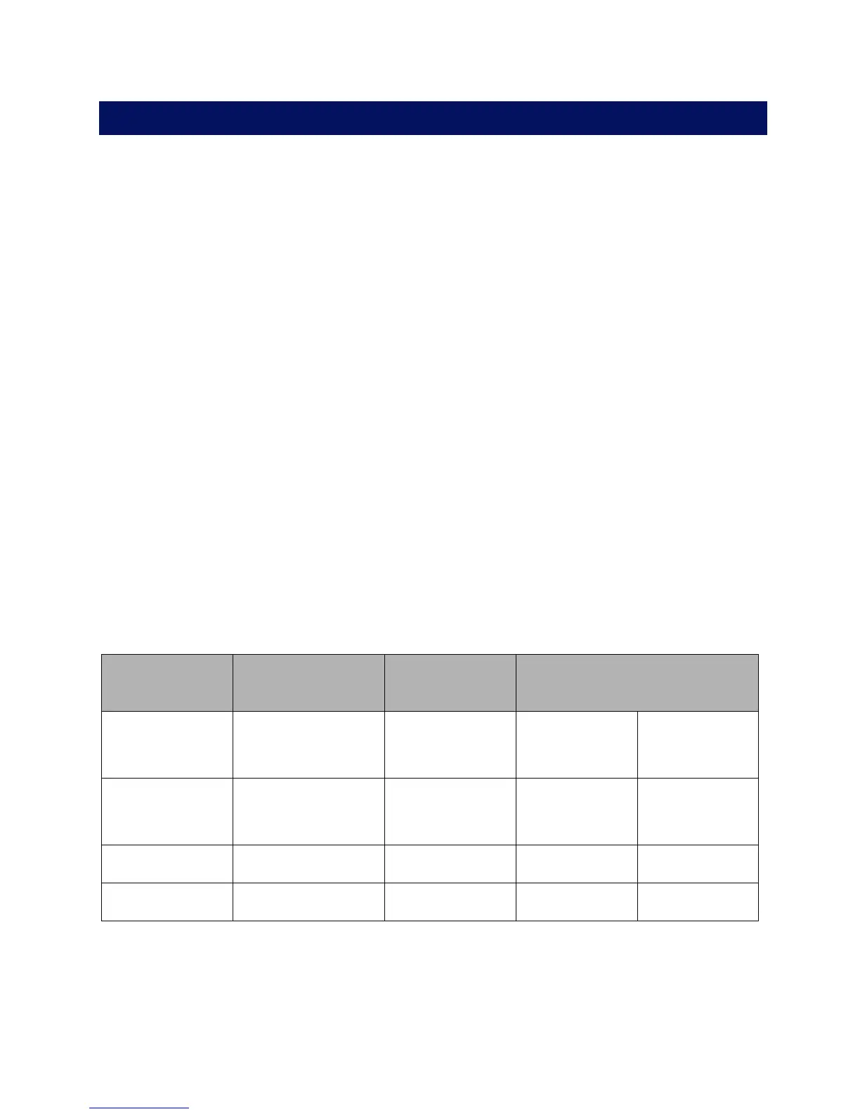
The following table lists the slice thickness, slice interval, kernel, and filter to use
for each scanner manufacturer:
Scanner
Manufacturer
Slice Thickness
(mm)
Slice Interval
(mm)
Kernel and Filter
GE™ 1.25 1.0
Kernel
Filter
Standard
Body
Philips™ 1.0 0.8
Kernel
Enhancement
C
0
Siemens™ 1.0 0.8 Kernel B31f
Toshiba™ 1.0 0.8 Kernel FC05