Do you have a question about the Ecleris VNG Plus and is the answer not in the manual?
Defines symbols used for warnings, cautions, and notes in the manual.
Outlines safety precautions for electrical shock, fire, ventilation, and approved accessories.
Explains various symbols used in the manual for information and safety.
Describes the purpose of VNG Plus in registering and analyzing involuntary eye movements.
Specifies that VNG Plus is for health professionals experienced in the technique.
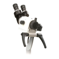
Explains how the VNG goggles use infrared light and mirrors to capture eye images.
States patients need adequate vision to follow targets for oculomotor tests.
Addresses patient's physical condition, especially neck/back injuries, for positional tests.
Details the importance of examining the outer and middle ear before caloric tests.
Explains how medications and alcohol can affect VNG test results.
Advises patients on preparation, including comfort, food intake, and transport.
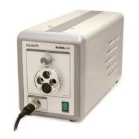
Lists essential parts like MainUnit, Goggles, Powercord, and User's Manual.
Details optional items such as sensors, T Bar, cushion mask, and footswitch.
Identifies the port for data connection on the front panel.
Describes the main power switch on the front panel.
Lists rear panel connections including AC INPUT, ULTRASOUND, VGA, USB, and Equipotential terminal.
Details connections for cameras and sensors to the junction box.
Identifies the sensor and cameras (Right/Left) on the goggles.
States that only authorized physicians can operate the system.
Guides users on starting the VNG Plus after installation.
Explains how to launch the VNG Plus software from the desktop or start menu.
Describes how to activate audio and video help for tests.
Details the process of entering patient name, chart number, DOB, and demographic data.
Guides on identifying a session, entering clinic data, and selecting a protocol.
Covers starting new tests and opening existing files or sessions.
Mentions accessing print reports as an additional function.
Explains resuming sessions, importing, and exporting tests.
Describes the purpose of toolbar buttons like Shut down, Setup, Support, and Start session.
Details how to define the sequence of tests within a custom protocol.
Explains how to view specific parameters for each test in a protocol.
Describes the function of Home, Back, Go To, and Next buttons in protocol screens.
Explains the DHI as a 25-item questionnaire measuring self-perceived disability from dizziness.
Guides on how to answer questions using buttons or keyboard input.
Outlines the function of Home, Back, and Go To buttons for the DHI screen.
Explains the importance of patient distance for visual stimulation geometry.
Notes that VNG Plus supports various screen geometries requiring configuration.
Describes the function of Home, Back, Go To, and Next buttons in the assistant interface.
Instructs on fitting the goggles securely to the patient's head for optimal performance.
Emphasizes centering the pupil for nystagmus assessment and image quality.
Suggests placing the junction box on the patient's arm to avoid wire tangles.
Explains the function of Home, Back, Go To, and Next buttons on the goggles screen.
Explains the need to select which eye's record to analyze, as only one can be processed at a time.
Warns that changing the eye for analysis after a session will lose previous analysis.
Describes the function of Home, Back, Go To, and Next buttons for eye selection.
Explains standard calibration as an allowance for correction factor, not amplitude.
Lists tests for which calibration is recommended, including Gaze, Saccades, OKN, etc.
Notes that VHIT and AHR tests require specific head movement calibration.
Guides the user to have the patient fixate on target points during calibration.
Explains the checkpoint as a quick verification after initial calibration.
Describes color indicators (green, yellow, red) for pupil detection status.
Details the function of Home, Back, Go To, and Next buttons during calibration.
Guides the patient to look at the target and describes progress bar status.
Outlines the function of Home, Back, Go To, and Next buttons for head calibration.
Explains patient-driven head movements synchronized with a metronome for AHR calibration.
Describes examiner-guided passive head movements for VHIT calibration.
Explains how eye position/velocity is graphically displayed against time.
Illustrates the display of saccadic test data, including eye position and stimulus.
Describes warnings for subject position, head angles, and head tilt during data acquisition.
Guides on accessing test instructions, recommendations, and video demonstrations.
Mentions the Back button for navigating the demo screen.
Details how to start recording a test and the options for stopping it.
Explains the utility of the foot switch for hands-free operation during tests.
Describes performing spontaneous nystagmus tests with vision occluded.
Details how to perform gaze testing and associated warnings.
Explains performing saccades tests and associated warnings.
Guides on performing smooth pursuit tests and associated warnings.
Describes performing OKN tests and associated warnings.
Explains performing AHR tests and the need for a loudspeaker.
Details performing vHIT tests and associated warnings.
Guides on performing supine position tests and associated warnings.
Describes the Dix Hallpike maneuver involving head turning and supine positioning.
Explains the use of fixation suppression (covered goggles) during testing.
Details reclining patient position with head at a 30° angle for caloric stimulation.
Lists standard caloric stimulation protocols (LW, RW, LC, RC).
Emphasizes the importance of examining ears before performing caloric tests.
Instructs patients to fixate on a red light to reduce induced nystagmus.
Describes the display of test data within the analysis window.
Explains how to choose specific tests or sub-tests from a dropdown menu.
Details how to interpret alarm status indicators (Excellent, Good, Poor).
Explains how to review video and alarm registers simultaneously.
Describes tools like Zoom, Mark, Edit, and Graphical Analysis for data review.
Covers viewing all test information and using zoom options for detailed data.
Explains spontaneous nystagmus may indicate central or peripheral pathology.
Lists parameters like Nystagmus Count and SPV for analysis.
Details how gaze tests document inability to maintain static eye position.
Lists parameters like Nystagmus Count and SPV for gaze analysis.
Notes that patient cooperation and visual acuity influence saccade test results.
Details parameters like Velocity, Latency, and Accuracy for saccades.
Explains the system's role in tracking targets and factors affecting results.
Mentions Gain, Phase, and THD as key factors for interpretation.
Describes how patients track multiple stimuli at programmed rates.
Highlights Gain and Asymmetry for OKN interpretation.
Explains AHR tests vestibular function during active head rotation.
Lists Gain, Phase, and THD of the vestibular ocular reflex.
States vHIT is useful for assessing semicircular canal deficits.
Details parameters like Velocity, Gain, and Acceleration for vHIT analysis.
Notes passive calibration results are shown graphically.
Lists parameters like speed and gain for passive head calibration.
Explains supine tests diagnose BPPV and other vestibular complaints.
Lists parameters calculated for horizontal channel in supine positions.
Highlights Dix Hallpike's importance in diagnosing BPPV and vestibular complaints.
Lists parameters calculated for horizontal channel on both sides.
Explains caloric tests evaluate peripheral end organ viability via stimulation.
Details parameters like preponderance, beats, and velocity for caloric analysis.
Provides the formula for calculating the Fixation Index (FI).
Describes managing report printing and configurations for various printers.
Explains how to preview records and analysis before printing.
Details options for printing directly to a printer or saving as a PDF.
Outlines functions like printing current page, all pages, and returning to the report window.
Lists key factors influencing nystagmus analysis accuracy.
Identifies image quality, eye detection, patient cooperation, and calibration as factors.
Explains how the software detects nystagmus beats and calculates parameters.
Describes overriding automatic analysis by adding or deleting nystagmus marks.
Explains analysis by separating rightward and leftward impulses.
Guides on adjusting sensitivity for automatic discarding of impulses.
Details how to manually discard impulses based on characteristics.
Describes accessing the test summary and selecting tests for export to external media.
Explains publishing tests (data, report, videos) on the Endodigiweb.com website.
Guides users on registering to create an ENDODIGIWEB account.
Details options for Public or Private access to published content.
Covers adding titles, abstracts, and inviting contacts to view publications.
Describes adding new contacts or importing from address books.
Confirms publication readiness after upload and provides upload success message.
Explains editing lists for patient records, doctors, administrators, and clinics.
Covers sorting, searching, and filtering stored sessions and patient data.
Guides on creating, adapting, and managing custom test protocols.
Details sorting by date/alphabetical order and using search tools for patients/sessions.
Explains the protocol tree structure and how to modify existing protocols.
Guides on creating new protocols and selecting tests by type/subtype.
Guides on accessing test instructions, recommendations, and video demonstrations.
Mentions the Back button for navigating the demo screen.
Introduces advanced settings grouped by characteristics for system configuration.
Describes toolbar buttons like Back, Default, and Save & Exit.
Explains selecting warning control levels (Soft, Normal, Hard).
Details setting up the visual stimulator for correct stimulation patterns.
Guides on inputting monitor dimensions for accurate stimulation.
Describes checking/unchecking items for printing and changing report settings.
Explains options like Auto Adjust, Graphics, Records, and Print Width.
Details setting the Time window for displaying or printing time plots.
Explains defining time offset and using scrollbars for chart views.
Guides on performing tests in user-defined sequences and controlling acquisition.
Lists various acquisition settings such as Alert Message, Test Instructions, and Render options.
Explains how to adjust detection algorithm sensitivity for specific tests.
Notes that pupil configuration requires technical support.
Lists detection parameters for pupil analysis.
Describes modifying the eye detection area to improve analysis results.
States that reprocessing is exclusively for technical support use.
Guides on accessing the support screen via the Support button.
Differentiates between Email Support and Remote Support.
Shows the screen displayed while waiting for a remote connection.
Displays the message when remote support is unavailable.
Emphasizes correct software installation before hardware installation.
Lists hardware (processor, memory, etc.) and software (OS, SDKs) requirements.
Details steps for installing the VNG Plus software using the pen drive.
Guides on selecting the installation language.
Recommends following default options during the installation screens.
Explains the automatic driver installation after file copying.
Describes the confirmation message upon successful driver installation.
Explains the automatic loading of USB Video Device and Data drivers.
Guides on verifying driver installation using Device Manager.
Details unpacking the device and warnings for receiver bar assembly.
Provides step-by-step instructions for assembling the receiver bar.
Guides on connecting the position sensor, cameras, and junction box.
Details connecting the monitor, PC, and power supply.
Instructs on powering on the device and verifying software installation.
Explains visual stimulation via digital images on a monitor.
Lists tests where the visual stimulator is used (Gaze, Smooth Pursuit, etc.).
Provides guidelines for eye height and ultrasonic sensor placement relative to the monitor.
Outlines precautions for cleaning the equipment to avoid damage.
Provides specific instructions for cleaning the console and receiver bar.
Details cleaning camera lenses, mirrors, frames, and mask straps.
Guides on replacing the goggle cushion pad.
Details the steps for opening the fuse compartment and replacing fuses.
Advises on disposing of removed parts according to regulations.
States that only qualified ECLERIS personnel should perform maintenance.
Cautions about using original packaging for transport and environmental conditions.
Addresses problems like the device not turning on or not being detected.
Solves issues related to image recording or display faults.
Troubleshoots problems with wireless switch detection or functionality.
Lists weight and dimensions for goggles and console.
Details power input, consumption, fuse type, and electrical protection class.
Specifies service, storage, and transport conditions.
Lists compliance with IEC60601 standards for medical electrical equipment.
Outlines system input, sampling, tracking range, and test capabilities.
Details specifications for video cameras and visual stimulus signals.
Defines abnormality thresholds for spontaneous nystagmus and bilateral gaze.
Lists abnormality thresholds for smooth pursuit gain, phase, and asymmetry.
Defines abnormality thresholds for saccade velocity, latency, and accuracy.
Lists abnormality thresholds for OKN gain and symmetry.
Defines abnormality thresholds for Dix-Hallpike test results.
Lists abnormality thresholds for caloric tests, including unilateral weakness and fixation index.
Explains the Freyss diagram for showing nystagmus intensity, symmetry, and preponderance.
Guides on interpreting the diagram's axes, lines, and intersection points.
Details formulas for calculating lateral symmetry and absolute preponderance.
Presents the DHI questionnaire structure and scoring system.
Provides manufacturer's declaration on electromagnetic emissions.
Details RF emissions for internal functioning, unlikely to cause interference.
Specifies suitability for use in establishments other than domestic premises.
States compliance with harmonic emissions standards.
Confirms compliance with flicker standards.
Provides manufacturer's declaration on electromagnetic immunity.
Details immunity levels for electrostatic discharge.
Specifies immunity levels for transients and rapid electric bursts.
Details immunity levels for surge voltages.
Lists immunity requirements for voltage fluctuations.
Specifies immunity levels for power frequency and magnetic fields.
Details immunity levels for conducted RF interference.
Specifies immunity levels for radiated RF interference.
| Category | Medical Equipment |
|---|---|
| Type | Videonystagmography (VNG) |
| Application | Vestibular and Balance Assessment |
| Components | Goggles with camera, Calibration unit, Control unit with software |
| Eye Tracking | Infrared video cameras for high-resolution eye movement recording |
| Software Features | Real-time analysis, Reporting |
| Compatibility | Windows-based PC |
| Calibration | Automated calibration procedure |
| Tests Supported | Spontaneous nystagmus, gaze, saccades, pursuit, optokinetic, positional, caloric |
| Data Export | PDF, CSV |