OPERATION
4.14
ThinPrep
®
Integrated Imager Operator’s Manual
Figure 4-14 Stage in Position for Specimen Adequacy Check
The system does not determine specimen adequacy; use your standard lab protocol. To estimate the cel-
lularity of the preparation in scantly cellular specimens, a specimen adequacy check can be performed.
In accordance with Bethesda criteria
1
, a minimum of 10 fields should be counted along a diameter of the
cell spot that includes the center. Dependent upon the microscope objective used, use the chart below
and count the average number of cells in each field.
Use the stage control knobs to traverse the cell spot.
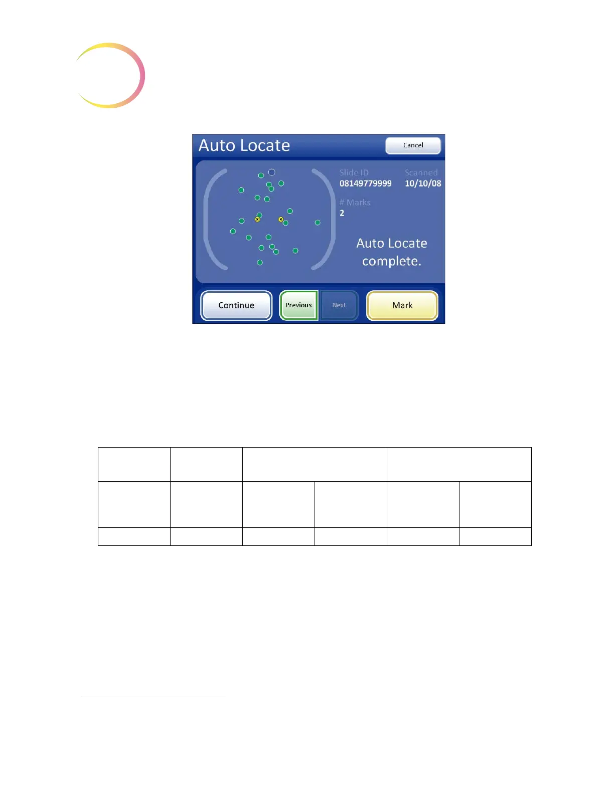
Once Auto Locate is complete, the operator may press the
Continue
button to proceed to:
• Auto Scan if any marks were made or further review is desired
• review electronic marks
• complete the review if no marks were made and no further review is desired (see page 4.18)
• press the
Cancel
button to cancel the review (No slide review data will be written to the data-
base.)
1. Nayar R, Wilbur DC. (eds). The Bethesda System for Reporting Cervical Cytology: Definitions, Criteria, and
Explanatory Notes. 3rd ed. Cham, Switzerland: Springer: 2015
FN 22 Eyepiece/
10X Objective
FN 22 Eyepiece/
40X Objective
PREP DIAM
(mm)
AREA
(mm
2
)
Total
Number of
Fields
Number of
Cells per Field
for 5,000 Total
Total
Number of
Fields
Number of
Cells per Field
for 5,000 Total
20 314.2 82.6 60.5 1322 3.8