3 Advanced use
KaVo Scan eXam One 21
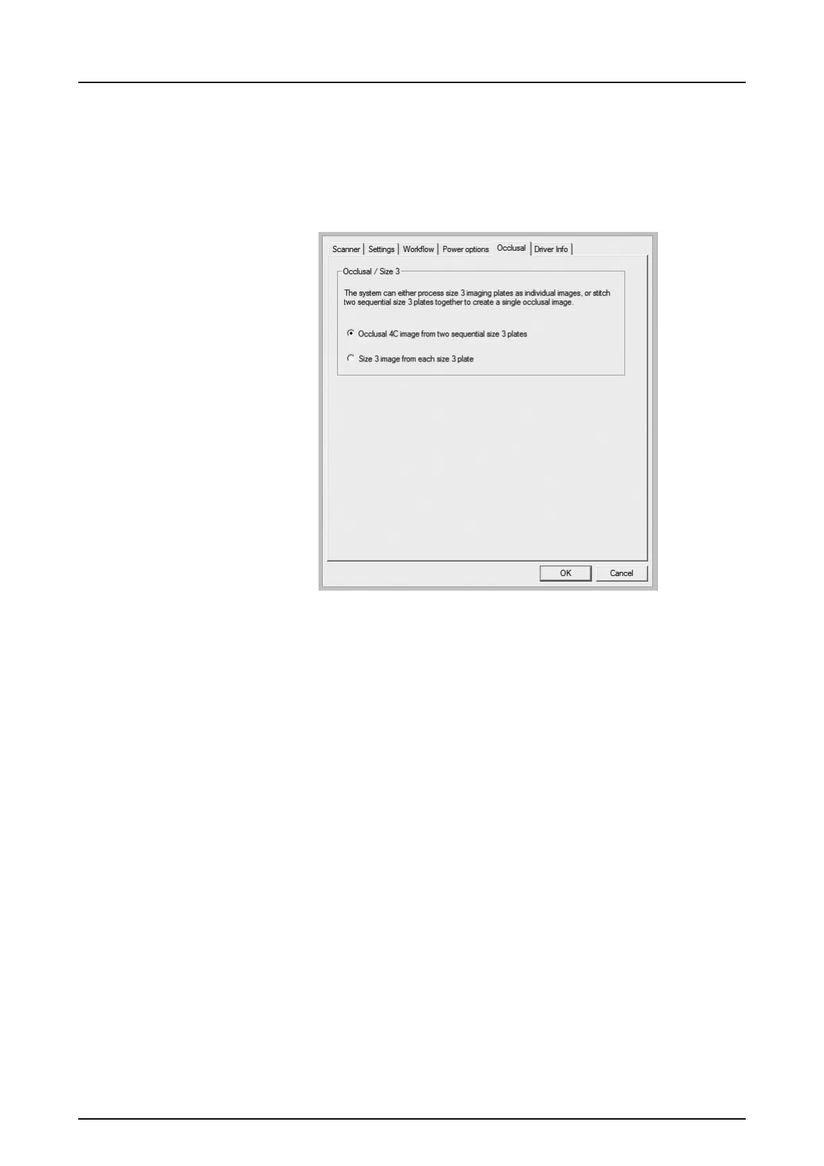
3.1.5 Occlusal 4C projection imaging
(not included in delivery)
To change Occlusal 4C projection imaging settings
select from the imaging application software unit
Setup / Occlusal page.
Occlusal 4C projection image is formed from two
sequential size 3 plates. Image plates are
processed separately and then stitched together to
form a single Occlusal 4C projection image.
Following text shortly describe how Occlusal 4C
projection image is taken. For more information
refer to instructions supplied with the Occlusal 4C
kit.
1. Place two size 3 imaging plates into their corre-
sponding protective covers.
2. Slide the two size 3 imaging plates and protec-
tive covers into the Occlusal 4C bite protector.
3. Insert the Occlusal 4C bite protector and imag-
ing plates into the Occlusal 4C hygiene bag.
4. Seal the bag. Place the sealed Occlusal 4C hy-
giene bag into the patient’s mouth and take an
exposure.
5. Remove the sealed Occlusal 4C hygiene bag
from the patient’s mouth. Open it.

 Loading...