3 Advanced use
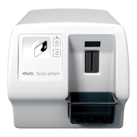
22 KaVo Scan eXam One
6. Remove each individual imaging plate from the
Occlusal 4C bite protector and process one at a
time.
7. Occlusal 4C image appear on the imaging
application software.
NOTICE! When you are in the Occlusal 4C mode it
is possible to temporarily override the mode and
process a single size 3 imaging plate. Insert the
size 3 imaging plate into the unit so that it can be
processed. When the insert second plate symbol
appears on the unit user interface press the start
key. This cancels the Occlusal 4C mode for this
operation and produce a single size 3 image.
Size 3 image mode from each size 3 plate allows
size 3 imaging plates to process as individual
imaging plates.
NOTICE! Due to Occlusal 4C projection imaging
geometry and imaging plate positioning, accurate
distance and angle measurements cannot be taken
from Occlusal 4C projection images.
3.2 Scan eXam™ One settings with
DTX Studio™ Core
You can view and change the device settings in DTX
Studio™ Core by following the steps below.
NOTICE! For most accurate and up to date
information, refer to DTX Studio™ Instructions for
Use.
1. Sign into DTX Studio™ Core with the same user
credentials as you use to login to DTX Studio™.
2. Select Manage devices menu.
 Loading...